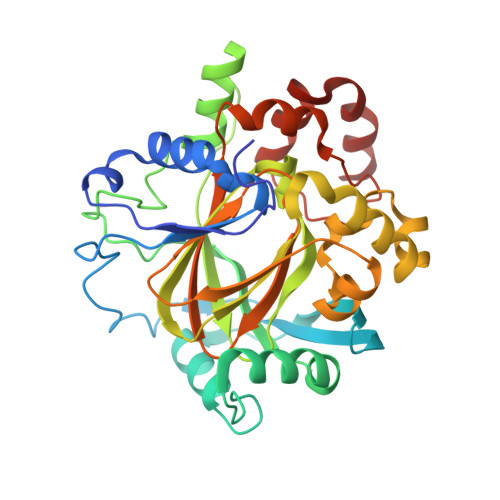
5FP8

Crystal structure of human KDM4D in complex with 3-4-methylthiophen-2- ylmethylaminopyridine-4-carboxylic acid


Experimental Data Snapshot

  • Method: X-RAY DIFFRACTION
  • Resolution: 1.98 Å
  • R-Value Free: 0.206 
  • R-Value Work: 0.159 
  • R-Value Observed: 0.162 

wwPDB Validation   3D Report Full Report


Ligand Structure Quality Assessment 


This is version 1.2 of the entry. See complete history


Literature

Cell Penetrant Inhibitors of the Kdm4 and Kdm5 Families of Histone Lysine Demethylases. 1. 3-Amino-4-Pyridine Carboxylate Derivatives.

Westaway, S.M.Preston, A.G.S.Barker, M.D.Brown, F.Brown, J.A.Campbell, M.Chung, C.Diallo, H.Douault, C.Drewes, G.Eagle, R.Gordon, L.Haslam, C.Hayhow, T.G.Humphreys, P.G.Joberty, G.Katso, R.Kruidenier, L.Leveridge, M.Liddle, J.Mosley, J.Muelbaier, M.Randle, R.Rioja, I.Rueger, A.Seal, G.A.Sheppard, R.J.Singh, O.Taylor, J.Thomas, P.Thomson, D.Wilson, D.M.Lee, K.Prinjha, R.K.

(2016) J Med Chem 59: 1357

  • DOI: https://doi.org/10.1021/acs.jmedchem.5b01537
  • Primary Citation of Related Structures:  
    5FP3, 5FP4, 5FP8, 5FP9, 5FPA, 5FPB

  • PubMed Abstract: 

    Optimization of KDM6B (JMJD3) HTS hit 12 led to the identification of 3-((furan-2-ylmethyl)amino)pyridine-4-carboxylic acid 34 and 3-(((3-methylthiophen-2-yl)methyl)amino)pyridine-4-carboxylic acid 39 that are inhibitors of the KDM4 (JMJD2) family of histone lysine demethylases. Compounds 34 and 39 possess activity, IC50 ≤ 100 nM, in KDM4 family biochemical (RFMS) assays with ≥ 50-fold selectivity against KDM6B and activity in a mechanistic KDM4C cell imaging assay (IC50 = 6-8 μM). Compounds 34 and 39 are also potent inhibitors of KDM5C (JARID1C) (RFMS IC50 = 100-125 nM).


  • Organizational Affiliation

    Epinova Discovery Performance Unit, Medicines Research Centre, GlaxoSmithKline R&D , Stevenage SG1 2NY, U.K.


Macromolecules
Find similar proteins by:  (by identity cutoff)  |  3D Structure
Entity ID: 1
MoleculeChains Sequence LengthOrganismDetailsImage
LYSINE-SPECIFIC DEMETHYLASE 4D334Homo sapiensMutation(s): 0 
EC: 1.14.11.66
UniProt & NIH Common Fund Data Resources
Find proteins for Q6B0I6 (Homo sapiens)
Explore Q6B0I6 
Go to UniProtKB:  Q6B0I6
PHAROS:  Q6B0I6
GTEx:  ENSG00000186280 
Entity Groups  
Sequence Clusters30% Identity50% Identity70% Identity90% Identity95% Identity100% Identity
UniProt GroupQ6B0I6
Sequence Annotations
Expand
  • Reference Sequence
Small Molecules
Ligands 4 Unique
IDChains Name / Formula / InChI Key2D Diagram3D Interactions
AUY
Query on AUY

Download Ideal Coordinates CCD File 
D [auth A]3-[(4-methylthiophen-2-yl)methylamino]pyridine-4-carboxylic acid
C12 H12 N2 O2 S
CXLKBDJGFBROEI-UHFFFAOYSA-N
SO4
Query on SO4

Download Ideal Coordinates CCD File 
E [auth A]
F [auth A]
G [auth A]
H [auth A]
I [auth A]
E [auth A],
F [auth A],
G [auth A],
H [auth A],
I [auth A],
J [auth A],
K [auth A]
SULFATE ION
O4 S
QAOWNCQODCNURD-UHFFFAOYSA-L
ZN
Query on ZN

Download Ideal Coordinates CCD File 
B [auth A]ZINC ION
Zn
PTFCDOFLOPIGGS-UHFFFAOYSA-N
CO
Query on CO

Download Ideal Coordinates CCD File 
C [auth A]COBALT (II) ION
Co
XLJKHNWPARRRJB-UHFFFAOYSA-N
Experimental Data & Validation

Experimental Data

  • Method: X-RAY DIFFRACTION
  • Resolution: 1.98 Å
  • R-Value Free: 0.206 
  • R-Value Work: 0.159 
  • R-Value Observed: 0.162 
  • Space Group: P 43 21 2
Unit Cell:
Length ( Å )Angle ( ˚ )
a = 71.286α = 90
b = 71.286β = 90
c = 151.054γ = 90
Software Package:
Software NamePurpose
REFMACrefinement
XDSdata reduction
SCALAdata scaling

Structure Validation

View Full Validation Report


Ligand Structure Quality Assessment 


Entry History 

Deposition Data

  • Released Date: 2016-01-27 
  • Deposition Author(s): Chung, C.

Revision History  (Full details and data files)

  • Version 1.0: 2016-01-27
    Type: Initial release
  • Version 1.1: 2016-03-09
    Changes: Database references
  • Version 1.2: 2024-05-08
    Changes: Data collection, Database references, Derived calculations, Other