5FOW | pdb_00005fow

HUMANISED MONOMERIC RADA IN COMPLEX WITH WHTA TETRAPEPTIDE


Experimental Data Snapshot

  • Method: X-RAY DIFFRACTION
  • Resolution: 1.80 Å
  • R-Value Free: 
    0.218 (Depositor), 0.219 (DCC) 
  • R-Value Work: 
    0.183 (Depositor), 0.185 (DCC) 
  • R-Value Observed: 
    0.185 (Depositor) 

Starting Model: experimental
View more details

wwPDB Validation 3D Report Full Report

Validation slider image for 5FOW

This is version 2.3 of the entry. See complete history

Literature

Structure Activity Relationship of the Peptide Binding Motif Mediating the Rad51:Brca2 Protein-Protein Interaction.

Scott, D.E.Marsh, M.Blundell, T.L.Abell, C.Hyvonen, M.

(2016) FEBS Lett 590: 1094

  • DOI: https://doi.org/10.1002/1873-3468.12139
  • Primary Citation Related Structures: 
    5FOT, 5FOU, 5FOV, 5FOW, 5FOX, 5FPK

  • PubMed Abstract: 

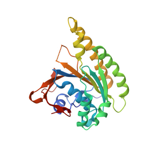
    RAD51 is a recombinase involved in the homologous recombination of double-strand breaks in DNA. RAD51 forms oligomers by binding to another molecule of RAD51 via an 'FxxA' motif, and the same recognition sequence is similarly utilised to bind BRCA2. We have tabulated the effects of mutation of this sequence, across a variety of experimental methods and from relevant mutations observed in the clinic. We use mutants of a tetrapeptide sequence to probe the binding interaction, using both isothermal titration calorimetry and X-ray crystallography. Where possible, comparison between our tetrapeptide mutational study and the previously reported mutations is made, discrepancies are discussed and the importance of secondary structure in interpreting alanine scanning and mutational data of this nature is considered.


  • Organizational Affiliation
    • Department of Chemistry, University of Cambridge, Cambridge, UK.

Macromolecule Content 

  • Total Structure Weight: 52.27 kDa 
  • Atom Count: 4,151 
  • Modeled Residue Count: 451 
  • Deposited Residue Count: 474 
  • Unique protein chains: 2

Macromolecules

Find similar proteins by:|  3D Structure
Entity ID: 1
MoleculeChains  Sequence LengthOrganismDetailsImage
DNA repair and recombination protein RadAA,
B [auth C]
231Pyrococcus furiosus DSM 3638Mutation(s): 5 
Gene Names: radAPF1926
UniProt
Find proteins for O74036 (Pyrococcus furiosus (strain ATCC 43587 / DSM 3638 / JCM 8422 / Vc1))
Explore O74036 
Go to UniProtKB:  O74036
Entity Groups
Sequence Clusters30% Identity50% Identity70% Identity90% Identity95% Identity100% Identity
UniProt GroupO74036
Sequence Annotations
Expand
Reference Sequence
Find similar proteins by:  Sequence   |   3D Structure  
Entity ID: 2
MoleculeChains  Sequence LengthOrganismDetailsImage
WHTA PEPTIDEC [auth E],
D [auth F]
6Homo sapiensMutation(s): 0 

Experimental Data & Validation

Experimental Data

  • Method: X-RAY DIFFRACTION
  • Resolution: 1.80 Å
  • R-Value Free:  0.218 (Depositor), 0.219 (DCC) 
  • R-Value Work:  0.183 (Depositor), 0.185 (DCC) 
  • R-Value Observed: 0.185 (Depositor) 
Space Group: P 1 21 1
Unit Cell:
Length ( Å )Angle ( ˚ )
a = 40.479α = 90
b = 87.598β = 92.37
c = 61.938γ = 90
Software Package:
Software NamePurpose
PHENIXrefinement
XDSdata reduction
XDSdata scaling
AMoREphasing

Structure Validation

View Full Validation Report


Entry History 

Deposition Data

Revision History  (Full details and data files)

  • Version 1.0: 2016-03-30
    Type: Initial release
  • Version 1.1: 2016-05-18
    Changes: Database references
  • Version 1.2: 2017-07-12
    Changes: Derived calculations
  • Version 2.0: 2019-06-19
    Changes: Advisory, Atomic model, Data collection, Database references, Derived calculations, Source and taxonomy, Structure summary
  • Version 2.1: 2019-08-21
    Changes: Data collection
  • Version 2.2: 2024-01-10
    Changes: Data collection, Database references, Other, Refinement description
  • Version 2.3: 2024-11-13
    Changes: Structure summary