5FOB | pdb_00005fob

Crystal Structure of Human Complement C3b in complex with Smallpox Inhibitor of Complement (SPICE)


Experimental Data Snapshot

  • Method: X-RAY DIFFRACTION
  • Resolution: 2.60 Å
  • R-Value Free: 
    0.230 (Depositor), 0.234 (DCC) 
  • R-Value Work: 
    0.198 (Depositor), 0.206 (DCC) 
  • R-Value Observed: 
    0.199 (Depositor) 

Starting Models: experimental
View more details

wwPDB Validation 3D Report Full Report

Validation slider image for 5FOB

This is version 2.2 of the entry. See complete history

Literature

Regulators of Complement Activity Mediate Inhibitory Mechanisms Through a Common C3B-Binding Mode.

Forneris, F.Wu, J.Xue, X.Ricklin, D.Lin, Z.Sfyroera, G.Tzekou, A.Volokhina, E.Granneman, J.C.Hauhart, R.Bertram, P.Liszewski, M.K.Atkinson, J.P.Lambris, J.D.Gros, P.

(2016) EMBO J 35: 1133

  • DOI: https://doi.org/10.15252/embj.201593673
  • Primary Citation Related Structures: 
    5FO7, 5FO8, 5FO9, 5FOA, 5FOB

  • PubMed Abstract: 

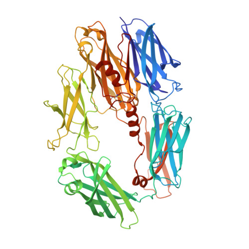
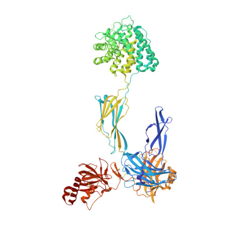
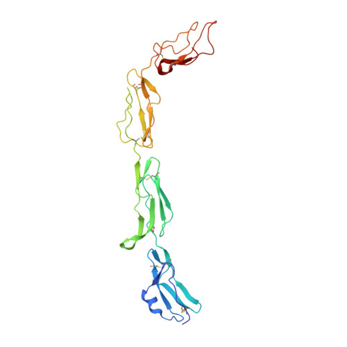
    Regulators of complement activation (RCA) inhibit complement-induced immune responses on healthy host tissues. We present crystal structures of human RCA (MCP, DAF, and CR1) and a smallpox virus homolog (SPICE) bound to complement component C3b. Our structural data reveal that up to four consecutive homologous CCP domains (i-iv), responsible for inhibition, bind in the same orientation and extended arrangement at a shared binding platform on C3b. Large sequence variations in CCP domains explain the diverse C3b-binding patterns, with limited or no contribution of some individual domains, while all regulators show extensive contacts with C3b for the domains at the third site. A variation of ~100° rotation around the longitudinal axis is observed for domains binding at the fourth site on C3b, without affecting the overall binding mode. The data suggest a common evolutionary origin for both inhibitory mechanisms, called decay acceleration and cofactor activity, with variable C3b binding through domains at sites ii, iii, and iv, and provide a framework for understanding RCA disease-related mutations and immune evasion.


  • Organizational Affiliation
    • Crystal and Structural Chemistry, Bijvoet Center for Biomolecular Research, Department of Chemistry, Faculty of Science Utrecht University, Utrecht, The Netherlands.

Macromolecule Content 

  • Total Structure Weight: 206.81 kDa 
  • Atom Count: 14,474 
  • Modeled Residue Count: 1,784 
  • Deposited Residue Count: 1,805 
  • Unique protein chains: 3

Macromolecules

Find similar proteins by:|  3D Structure
Entity ID: 1
MoleculeChains  Sequence LengthOrganismDetailsImage
COMPLEMENT C3 BETA CHAIN645Homo sapiensMutation(s): 0 
UniProt & NIH Common Fund Data Resources
Find proteins for P01024 (Homo sapiens)
Explore P01024 
Go to UniProtKB:  P01024
PHAROS:  P01024
GTEx:  ENSG00000125730 
Entity Groups
Sequence Clusters30% Identity50% Identity70% Identity90% Identity95% Identity100% Identity
UniProt GroupP01024
Glycosylation
Glycosylation Sites: 1Go to GlyGen: P01024-1
Sequence Annotations
Expand
Reference Sequence
Find similar proteins by:|  3D Structure
Entity ID: 2
MoleculeChains  Sequence LengthOrganismDetailsImage
COMPLEMENT C3B ALPHA' CHAIN915Homo sapiensMutation(s): 0 
UniProt & NIH Common Fund Data Resources
Find proteins for P01024 (Homo sapiens)
Explore P01024 
Go to UniProtKB:  P01024
PHAROS:  P01024
GTEx:  ENSG00000125730 
Entity Groups
Sequence Clusters30% Identity50% Identity70% Identity90% Identity95% Identity100% Identity
UniProt GroupP01024
Glycosylation
Glycosylation Sites: 1Go to GlyGen: P01024-1
Sequence Annotations
Expand
Reference Sequence
Find similar proteins by:|  3D Structure
Entity ID: 3
MoleculeChains  Sequence LengthOrganismDetailsImage
SMALLPOX INHIBITOR OF COMPLEMENT SPICE, D15L245Variola major virusMutation(s): 0 
UniProt
Find proteins for Q76U65 (Variola virus)
Explore Q76U65 
Go to UniProtKB:  Q76U65
Entity Groups
Sequence Clusters30% Identity50% Identity70% Identity90% Identity95% Identity100% Identity
UniProt GroupQ76U65
Sequence Annotations
Expand
Reference Sequence

Oligosaccharides

Help  
Entity ID: 4
MoleculeChains Length2D Diagram GlycosylationD Interactions
beta-D-mannopyranose-(1-4)-2-acetamido-2-deoxy-beta-D-glucopyranose-(1-4)-[alpha-L-fucopyranose-(1-6)]2-acetamido-2-deoxy-beta-D-glucopyranose
D
4N-Glycosylation
Glycosylation Resources
GlyTouCan: G32152BH
GlyCosmos: G32152BH
GlyGen: G32152BH
Entity ID: 5
MoleculeChains Length2D Diagram GlycosylationD Interactions
2-acetamido-2-deoxy-beta-D-glucopyranose-(1-4)-2-acetamido-2-deoxy-beta-D-glucopyranose
E
2N-Glycosylation
Glycosylation Resources
GlyTouCan: G42666HT
GlyCosmos: G42666HT
GlyGen: G42666HT

Small Molecules

Ligands 4 Unique
IDChains Name / Formula / InChI Key2D Diagram3D Interactions
IOD

Query on IOD


Download:Ideal Coordinates CCD File
IA [auth B]
JA [auth B]
KA [auth B]
LA [auth B]
R [auth A]
IA [auth B],
JA [auth B],
KA [auth B],
LA [auth B],
R [auth A],
S [auth A],
T [auth A]
IODIDE ION
I
XMBWDFGMSWQBCA-UHFFFAOYSA-M
GOL

Query on GOL


Download:Ideal Coordinates CCD File
AA [auth B]
BA [auth B]
CA [auth B]
DA [auth B]
EA [auth B]
AA [auth B],
BA [auth B],
CA [auth B],
DA [auth B],
EA [auth B],
FA [auth B],
G [auth A],
H [auth A],
I [auth A],
J [auth A],
K [auth A],
L [auth A],
M [auth A],
N [auth A],
NA [auth C],
O [auth A],
OA [auth C],
P [auth A],
U [auth B],
V [auth B],
W [auth B],
X [auth B],
Y [auth B],
Z [auth B]
GLYCEROL
C3 H8 O3
PEDCQBHIVMGVHV-UHFFFAOYSA-N
CL

Query on CL


Download:Ideal Coordinates CCD File
F [auth A],
GA [auth B],
MA [auth B],
PA [auth C],
QA [auth C]
CHLORIDE ION
Cl
VEXZGXHMUGYJMC-UHFFFAOYSA-M
NA

Query on NA


Download:Ideal Coordinates CCD File
HA [auth B],
Q [auth A]
SODIUM ION
Na
FKNQFGJONOIPTF-UHFFFAOYSA-N

Experimental Data & Validation

Experimental Data

  • Method: X-RAY DIFFRACTION
  • Resolution: 2.60 Å
  • R-Value Free:  0.230 (Depositor), 0.234 (DCC) 
  • R-Value Work:  0.198 (Depositor), 0.206 (DCC) 
  • R-Value Observed: 0.199 (Depositor) 
Space Group: P 1
Unit Cell:
Length ( Å )Angle ( ˚ )
a = 68.891α = 75.03
b = 83.311β = 76.2
c = 127.377γ = 68.5
Software Package:
Software NamePurpose
PHENIXrefinement
XDSdata reduction
Aimlessdata scaling
PHASERphasing

Structure Validation

View Full Validation Report


Entry History 

Deposition Data

Revision History  (Full details and data files)

  • Version 1.0: 2016-04-06
    Type: Initial release
  • Version 1.1: 2016-06-01
    Changes: Database references
  • Version 2.0: 2020-07-29
    Type: Remediation
    Reason: Carbohydrate remediation
    Changes: Advisory, Atomic model, Data collection, Derived calculations, Other, Structure summary
  • Version 2.1: 2024-01-10
    Changes: Data collection, Database references, Refinement description, Structure summary
  • Version 2.2: 2024-10-16
    Changes: Structure summary