Functional and Structural Characterization of a Potent Gh74 Endo-Xyloglucanase from the Soil Saprophyte Cellvibrio Japonicus Unravels the First Step of Xyloglucan Degradation.
Attia, M., Stepper, J., Davies, G.J., Brumer, H.(2016) FEBS J 283: 1701
- PubMed: 26929175
- DOI: https://doi.org/10.1111/febs.13696
- Primary Citation of Related Structures:
5FKQ, 5FKR, 5FKS, 5FKT - PubMed Abstract:
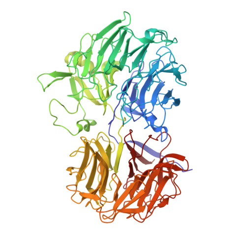
The heteropolysaccharide xyloglucan (XyG) comprises up to one-quarter of the total carbohydrate content of terrestrial plant cell walls and, as such, represents a significant reservoir in the global carbon cycle. The complex composition of XyG requires a consortium of backbone-cleaving endo-xyloglucanases and side-chain cleaving exo-glycosidases for complete saccharification. The biochemical basis for XyG utilization by the model Gram-negative soil saprophytic bacterium Cellvibrio japonicus is incompletely understood, despite the recent characterization of associated side-chain cleaving exo-glycosidases. We present a detailed functional and structural characterization of a multimodular enzyme encoded by gene locus CJA_2477. The CJA_2477 gene product comprises an N-terminal glycoside hydrolase family 74 (GH74) endo-xyloglucanase module in train with two carbohydrate-binding modules (CBMs) from families 10 and 2 (CBM10 and CBM2). The GH74 catalytic domain generates Glc4 -based xylogluco-oligosaccharide (XyGO) substrates for downstream enzymes through an endo-dissociative mode of action. X-ray crystallography of the GH74 module, alone and in complex with XyGO products spanning the entire active site, revealed a broad substrate-binding cleft specifically adapted to XyG recognition, which is composed of two seven-bladed propeller domains characteristic of the GH74 family. The appended CBM10 and CBM2 members notably did not bind XyG, nor other soluble polysaccharides, and instead were specific cellulose-binding modules. Taken together, these data shed light on the first step of xyloglucan utilization by C. japonicus and expand the repertoire of GHs and CBMs for selective biomass analysis and utilization. Structural data have been deposited in the RCSB protein database under the Protein Data Bank codes: 5FKR, 5FKS, 5FKT and 5FKQ.
Organizational Affiliation:
Michael Smith Laboratories and Department of Chemistry, University of British Columbia, Vancouver, Canada.


