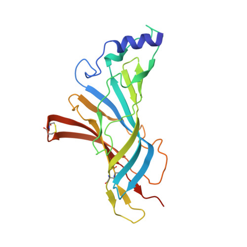
5FJV

Crystal structure of the extracellular domain of alpha2 nicotinic acetylcholine receptor in pentameric assembly


Experimental Data Snapshot

  • Method: X-RAY DIFFRACTION
  • Resolution: 3.20 Å
  • R-Value Free: 0.308 
  • R-Value Work: 0.224 
  • R-Value Observed: 0.228 

Starting Model: experimental
View more details

wwPDB Validation   3D Report Full Report


Ligand Structure Quality Assessment 


This is version 1.4 of the entry. See complete history


Literature

Crystal Structure of a Human Neuronal Nachr Extracellular Domain in Pentameric Assembly: Ligand-Bound Alpah2 Homopentamer.

Kouvatsos, N.Giastas, P.Chroni-Tzartou, D.Poulopoulou, C.Tzartos, S.J.

(2016) Proc Natl Acad Sci U S A 113: 9635

  • DOI: https://doi.org/10.1073/pnas.1602619113
  • Primary Citation of Related Structures:  
    5FJV

  • PubMed Abstract: 

    In this study we report the X-ray crystal structure of the extracellular domain (ECD) of the human neuronal α2 nicotinic acetylcholine receptor (nAChR) subunit in complex with the agonist epibatidine at 3.2 Å. Interestingly, α2 was crystallized as a pentamer, revealing the intersubunit interactions in a wild type neuronal nAChR ECD and the full ligand binding pocket conferred by two adjacent α subunits. The pentameric assembly presents the conserved structural scaffold observed in homologous proteins, as well as distinctive features, providing unique structural information of the binding site between principal and complementary faces. Structure-guided mutagenesis and electrophysiological data confirmed the presence of the α2(+)/α2(-) binding site on the heteromeric low sensitivity α2β2 nAChR and validated the functional importance of specific residues in α2 and β2 nAChR subunits. Given the pathological importance of the α2 nAChR subunit and the high sequence identity with α4 (78%) and other neuronal nAChR subunits, our findings offer valuable information for modeling several nAChRs and ultimately for structure-based design of subtype specific drugs against the nAChR associated diseases.


  • Organizational Affiliation

    Department of Neurobiology, Hellenic Pasteur Institute, 11521 Athens, Greece; nkouvatsos@pasteur.gr tzartos@pasteur.gr.


Macromolecules
Find similar proteins by:  (by identity cutoff)  |  3D Structure
Entity ID: 1
MoleculeChains Sequence LengthOrganismDetailsImage
NEURONAL ACETYLCHOLINE RECEPTOR SUBUNIT ALPHA-2
A, B, C, D, E
213Homo sapiensMutation(s): 0 
UniProt & NIH Common Fund Data Resources
Find proteins for Q15822 (Homo sapiens)
Explore Q15822 
Go to UniProtKB:  Q15822
PHAROS:  Q15822
GTEx:  ENSG00000120903 
Entity Groups  
Sequence Clusters30% Identity50% Identity70% Identity90% Identity95% Identity100% Identity
UniProt GroupQ15822
Sequence Annotations
Expand
  • Reference Sequence
Experimental Data & Validation

Experimental Data

  • Method: X-RAY DIFFRACTION
  • Resolution: 3.20 Å
  • R-Value Free: 0.308 
  • R-Value Work: 0.224 
  • R-Value Observed: 0.228 
  • Space Group: P 21 21 21
Unit Cell:
Length ( Å )Angle ( ˚ )
a = 101.524α = 90
b = 116.559β = 90
c = 129.546γ = 90
Software Package:
Software NamePurpose
PHENIXrefinement
XDSdata reduction
XDSdata scaling
PHASERphasing

Structure Validation

View Full Validation Report


Ligand Structure Quality Assessment 


Entry History 

Deposition Data

Revision History  (Full details and data files)

  • Version 1.0: 2016-08-10
    Type: Initial release
  • Version 1.1: 2016-08-17
    Changes: Database references
  • Version 1.2: 2016-09-28
    Changes: Database references
  • Version 1.3: 2024-01-10
    Changes: Data collection, Database references, Derived calculations, Other, Refinement description
  • Version 1.4: 2024-10-23
    Changes: Structure summary