5FJ7 | pdb_00005fj7

Structure of the P2 polymerase inside in vitro assembled bacteriophage phi6 polymerase complex, with P1 included


Experimental Data Snapshot

  • Method: ELECTRON MICROSCOPY
  • Resolution: 7.90 Å
  • Aggregation State: PARTICLE 
  • Reconstruction Method: SINGLE PARTICLE 

Starting Models: experimental
View more details

wwPDB Validation 3D Report Full Report

Validation slider image for 5FJ7

This is version 1.4 of the entry. See complete history

Literature

Localized Reconstruction of Subunits from Electron Cryomicroscopy Images of Macromolecular Complexes.

Ilca, S.L.Kotecha, A.Sun, X.Poranen, M.M.Stuart, D.I.Huiskonen, J.T.

(2015) Nat Commun 6: 8843

  • DOI: https://doi.org/10.1038/ncomms9843
  • Primary Citation Related Structures: 
    5FJ5, 5FJ6, 5FJ7

  • PubMed Abstract: 

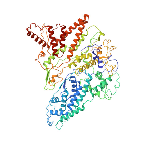
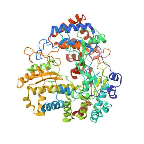
    Electron cryomicroscopy can yield near-atomic resolution structures of highly ordered macromolecular complexes. Often however some subunits bind in a flexible manner, have different symmetry from the rest of the complex, or are present in sub-stoichiometric amounts, limiting the attainable resolution. Here we report a general method for the localized three-dimensional reconstruction of such subunits. After determining the particle orientations, local areas corresponding to the subunits can be extracted and treated as single particles. We demonstrate the method using three examples including a flexible assembly and complexes harbouring subunits with either partial occupancy or mismatched symmetry. Most notably, the method allows accurate fitting of the monomeric RNA-dependent RNA polymerase bound at the threefold axis of symmetry inside a viral capsid, revealing for the first time its exact orientation and interactions with the capsid proteins. Localized reconstruction is expected to provide novel biological insights in a range of challenging biological systems.


  • Organizational Affiliation
    • Division of Structural Biology, Wellcome Trust Centre for Human Genetics, University of Oxford, Roosevelt Drive, Oxford OX3 7BN, UK.

Macromolecule Content 

  • Total Structure Weight: 243.29 kDa 
  • Atom Count: 17,106 
  • Modeled Residue Count: 2,186 
  • Deposited Residue Count: 2,186 
  • Unique protein chains: 2

Macromolecules

Find similar proteins by:|  3D Structure
Entity ID: 1
MoleculeChains  Sequence LengthOrganismDetailsImage
MAJOR INNER PROTEIN P1
A, B
761Cystovirus phi6Mutation(s): 0 
UniProt
Find proteins for P11126 (Pseudomonas phage phi6)
Explore P11126 
Go to UniProtKB:  P11126
Entity Groups
Sequence Clusters30% Identity50% Identity70% Identity90% Identity95% Identity100% Identity
UniProt GroupP11126
Sequence Annotations
Expand
Reference Sequence
Find similar proteins by:|  3D Structure
Entity ID: 2
MoleculeChains  Sequence LengthOrganismDetailsImage
RNA-DIRECTED RNA POLYMERASE664Cystovirus phi6Mutation(s): 0 
EC: 2.7.7.48
UniProt
Find proteins for P11124 (Pseudomonas phage phi6)
Explore P11124 
Go to UniProtKB:  P11124
Entity Groups
Sequence Clusters30% Identity50% Identity70% Identity90% Identity95% Identity100% Identity
UniProt GroupP11124
Sequence Annotations
Expand
Reference Sequence

Small Molecules

Ligands 1 Unique
IDChains Name / Formula / InChI Key2D Diagram3D Interactions
MN

Query on MN


Download:Ideal Coordinates CCD File
D [auth C]MANGANESE (II) ION
Mn
WAEMQWOKJMHJLA-UHFFFAOYSA-N

Experimental Data & Validation

Experimental Data

  • Method: ELECTRON MICROSCOPY
  • Resolution: 7.90 Å
  • Aggregation State: PARTICLE 
  • Reconstruction Method: SINGLE PARTICLE 
EM Software:
TaskSoftware PackageVersion
RECONSTRUCTIONRELION

Structure Validation

View Full Validation Report


Entry History 

Deposition Data

Revision History  (Full details and data files)

  • Version 1.0: 2015-11-04
    Type: Initial release
  • Version 1.1: 2015-11-18
    Changes: Database references
  • Version 1.2: 2017-08-02
    Changes: Data collection, Refinement description
  • Version 1.3: 2019-11-20
    Changes: Derived calculations
  • Version 1.4: 2024-05-08
    Changes: Data collection, Database references, Derived calculations, Refinement description