5FIY | pdb_00005fiy

crystal structure of coiled coil domain of PAWR


Experimental Data Snapshot

  • Method: X-RAY DIFFRACTION
  • Resolution: 3.00 Å
  • R-Value Free: 
    0.294 (Depositor), 0.312 (DCC) 
  • R-Value Work: 
    0.245 (Depositor), 0.269 (DCC) 
  • R-Value Observed: 
    0.250 (Depositor) 

Starting Model: other
View more details

wwPDB Validation 3D Report Full Report

Validation slider image for 5FIY

This is version 1.4 of the entry. See complete history

Literature

Structural basis for the regulatory interactions of proapoptotic Par-4.

Tiruttani Subhramanyam, U.K.Kubicek, J.Eidhoff, U.B.Labahn, J.

(2017) Cell Death Differ 24: 1540-1547

  • DOI: https://doi.org/10.1038/cdd.2017.76
  • Primary Citation Related Structures: 
    5FIY

  • PubMed Abstract: 

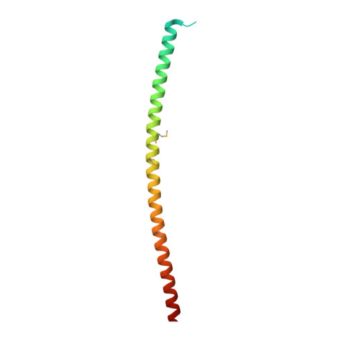
    Par-4 is a unique proapoptotic protein with the ability to induce apoptosis selectively in cancer cells. The X-ray crystal structure of the C-terminal domain of Par-4 (Par-4 CC ), which regulates its apoptotic function, was obtained by MAD phasing. Par-4 homodimerizes by forming a parallel coiled-coil structure. The N-terminal half of Par-4 CC contains the homodimerization subdomain. This structure includes a nuclear export signal (Par-4 NES ) sequence, which is masked upon dimerization indicating a potential mechanism for nuclear localization. The heteromeric-interaction models specifically showed that charge interaction is an important factor in the stability of heteromers of the C-terminal leucine zipper subdomain of Par-4 (Par-4 LZ ). These heteromer models also displayed NES masking capacity and therefore the ability to influence intracellular localization.


  • Organizational Affiliation
    • Centre for Structural Systems Biology (CSSB), Hamburg, Germany.

Macromolecule Content 

  • Total Structure Weight: 87.51 kDa 
  • Atom Count: 3,958 
  • Modeled Residue Count: 469 
  • Deposited Residue Count: 728 
  • Unique protein chains: 1

Macromolecules

Find similar proteins by:|  3D Structure
Entity ID: 1
MoleculeChains  Sequence LengthOrganismDetailsImage
PRKC APOPTOSIS WT1 REGULATOR PROTEIN
A, B, C, D, E
A, B, C, D, E, F, G
104Rattus norvegicusMutation(s): 0 
UniProt
Find proteins for Q62627 (Rattus norvegicus)
Explore Q62627 
Go to UniProtKB:  Q62627
Entity Groups
Sequence Clusters30% Identity50% Identity70% Identity90% Identity95% Identity100% Identity
UniProt GroupQ62627
Sequence Annotations
Expand
Reference Sequence

Small Molecules

Ligands 1 Unique
IDChains Name / Formula / InChI Key2D Diagram3D Interactions
K

Query on K


Download:Ideal Coordinates CCD File
H [auth A]
I [auth A]
J [auth A]
K [auth B]
L [auth B]
H [auth A],
I [auth A],
J [auth A],
K [auth B],
L [auth B],
M [auth B],
N [auth C],
O [auth C],
P [auth C],
Q [auth C],
R [auth D],
S [auth D],
T [auth D],
U [auth E],
V [auth E],
W [auth E],
X [auth F],
Y [auth F]
POTASSIUM ION
K
NPYPAHLBTDXSSS-UHFFFAOYSA-N
Modified Residues  1 Unique
IDChains TypeFormula2D DiagramParent
MSE
Query on MSE
A, B, C, D, E
A, B, C, D, E, F, G
L-PEPTIDE LINKINGC5 H11 N O2 SeMET

Experimental Data & Validation

Experimental Data

  • Method: X-RAY DIFFRACTION
  • Resolution: 3.00 Å
  • R-Value Free:  0.294 (Depositor), 0.312 (DCC) 
  • R-Value Work:  0.245 (Depositor), 0.269 (DCC) 
  • R-Value Observed: 0.250 (Depositor) 
Space Group: P 43 21 2
Unit Cell:
Length ( Å )Angle ( ˚ )
a = 114.99α = 90
b = 114.99β = 90
c = 121.81γ = 90
Software Package:
Software NamePurpose
PHENIXrefinement
MOSFLMdata reduction
Aimlessdata scaling
AutoSolphasing

Structure Validation

View Full Validation Report


Entry History 

Deposition Data

Revision History  (Full details and data files)

  • Version 1.0: 2016-11-16
    Type: Initial release
  • Version 1.1: 2017-08-30
    Changes: Database references
  • Version 1.2: 2017-12-06
    Changes: Database references
  • Version 1.3: 2024-05-01
    Changes: Data collection, Database references, Derived calculations, Other, Refinement description
  • Version 1.4: 2024-11-20
    Changes: Structure summary