5FHN | pdb_00005fhn

Crystal structure of the GluA2 ligand-binding domain (S1S2J) in complex with (S)-2-Amino-3-(5-(2-(3-methylbenzyl)-2H-tetrazol-5-yl)-3-hydroxyisoxazol-4-yl)propanoic acid at 1.6 A resolution


Experimental Data Snapshot

  • Method: X-RAY DIFFRACTION
  • Resolution: 1.60 Å
  • R-Value Free: 
    0.186 (Depositor), 0.189 (DCC) 
  • R-Value Work: 
    0.156 (Depositor), 0.155 (DCC) 
  • R-Value Observed: 
    0.157 (Depositor) 

Starting Model: experimental
View more details

wwPDB Validation 3D Report Full Report

Validation slider image for 5FHN

Ligand Structure Quality Assessment 


This is version 1.4 of the entry. See complete history

Literature

Tweaking Subtype Selectivity and Agonist Efficacy at (S)-2-Amino-3-(3-hydroxy-5-methyl-isoxazol-4-yl)propionic acid (AMPA) Receptors in a Small Series of BnTetAMPA Analogues.

Wang, S.Y.Larsen, Y.Navarrete, C.V.Jensen, A.A.Nielsen, B.Al-Musaed, A.Frydenvang, K.Kastrup, J.S.Pickering, D.S.Clausen, R.P.

(2016) J Med Chem 59: 2244-2254

  • DOI: https://doi.org/10.1021/acs.jmedchem.5b01982
  • Primary Citation Related Structures: 
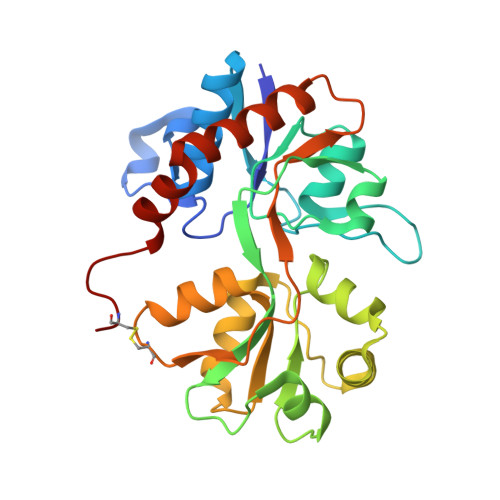
    5FHM, 5FHN, 5FHO

  • PubMed Abstract: 

    A series of analogues of the (S)-2-Amino-3-(3-hydroxy-5-methyl-isoxazol-4-yl)propionic acid (AMPA) receptor agonist BnTetAMPA (5b) were synthesized and characterized pharmacologically in radioligand binding assays at native and cloned AMPA receptors and functionally by two-electrode voltage clamp electrophysiology at the four homomeric AMPA receptors expressed in Xenopus laevis oocytes. The analogues 6 and 7 exhibit very different pharmacological profiles with binding affinity preference for the subtypes GluA1 and GluA3, respectively. X-ray crystal structures of three ligands (6, 7, and 8) in complex with the agonist binding domain (ABD) of GluA2 show that they induce full domain closure despite their low agonist efficacies. Trp767 in GluA2 ABD could be an important determinant for partial agonism of this compound series at AMPA receptors, since agonist efficacy also correlated with the location of the Trp767 side chain.


  • Organizational Affiliation
    • Department of Drug Design and Pharmacology, Faculty of Health and Medical Sciences, University of Copenhagen , Universitetsparken 2, DK 2100 Copenhagen, Denmark.

Macromolecule Content 

  • Total Structure Weight: 30.02 kDa 
  • Atom Count: 2,375 
  • Modeled Residue Count: 258 
  • Deposited Residue Count: 264 
  • Unique protein chains: 1

Macromolecules

Find similar proteins by:|  3D Structure
Entity ID: 1
MoleculeChains  Sequence LengthOrganismDetailsImage
Glutamate receptor 2,Glutamate receptor 2264Rattus norvegicusMutation(s): 0 
Gene Names: Gria2Glur2
UniProt
Find proteins for P19491 (Rattus norvegicus)
Explore P19491 
Go to UniProtKB:  P19491
Entity Groups
Sequence Clusters30% Identity50% Identity70% Identity90% Identity95% Identity100% Identity
UniProt GroupP19491
Sequence Annotations
Expand
Reference Sequence

Small Molecules

Ligands 5 Unique
IDChains Name / Formula / InChI Key2D Diagram3D Interactions
5XO

Query on 5XO


Download:Ideal Coordinates CCD File
F [auth A](S)-2-Amino-3-(5-(2-(3-methylbenzyl)-2H-tetrazol-5-yl)-3-hydroxyisoxazol-4-yl)propanoic acid
C15 H16 N6 O4
VGTSUUCRWSMODJ-NSHDSACASA-N
SO4

Query on SO4


Download:Ideal Coordinates CCD File
B [auth A]SULFATE ION
O4 S
QAOWNCQODCNURD-UHFFFAOYSA-L
GOL

Query on GOL


Download:Ideal Coordinates CCD File
C [auth A],
D [auth A]
GLYCEROL
C3 H8 O3
PEDCQBHIVMGVHV-UHFFFAOYSA-N
EDO

Query on EDO


Download:Ideal Coordinates CCD File
G [auth A]1,2-ETHANEDIOL
C2 H6 O2
LYCAIKOWRPUZTN-UHFFFAOYSA-N
ACT

Query on ACT


Download:Ideal Coordinates CCD File
E [auth A]ACETATE ION
C2 H3 O2
QTBSBXVTEAMEQO-UHFFFAOYSA-M

Experimental Data & Validation

Experimental Data

  • Method: X-RAY DIFFRACTION
  • Resolution: 1.60 Å
  • R-Value Free:  0.186 (Depositor), 0.189 (DCC) 
  • R-Value Work:  0.156 (Depositor), 0.155 (DCC) 
  • R-Value Observed: 0.157 (Depositor) 
Space Group: C 1 2 1
Unit Cell:
Length ( Å )Angle ( ˚ )
a = 137.11α = 90
b = 46.38β = 92.21
c = 46.03γ = 90
Software Package:
Software NamePurpose
MOSFLMdata reduction
SCALAdata scaling
PHENIXrefinement
PDB_EXTRACTdata extraction
PHASERphasing
Cootmodel building

Structure Validation

View Full Validation Report


Ligand Structure Quality Assessment 


Entry History 

Deposition Data

Revision History  (Full details and data files)

  • Version 1.0: 2016-03-02
    Type: Initial release
  • Version 1.1: 2018-01-17
    Changes: Data collection
  • Version 1.2: 2021-03-31
    Changes: Derived calculations
  • Version 1.3: 2024-01-10
    Changes: Data collection, Database references, Refinement description
  • Version 1.4: 2024-10-16
    Changes: Structure summary