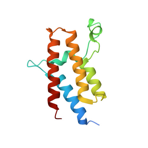
5FH7

Crystal structure of the fifth bromodomain of human PB1 in complex with compound 18


Experimental Data Snapshot

  • Method: X-RAY DIFFRACTION
  • Resolution: 1.47 Å
  • R-Value Free: 0.208 
  • R-Value Work: 0.187 
  • R-Value Observed: 0.188 

Starting Model: experimental
View more details

wwPDB Validation   3D Report Full Report


Ligand Structure Quality Assessment 


This is version 1.2 of the entry. See complete history


Literature

Identification and Development of 2,3-Dihydropyrrolo[1,2-a]quinazolin-5(1H)-one Inhibitors Targeting Bromodomains within the Switch/Sucrose Nonfermenting Complex.

Sutherell, C.L.Tallant, C.Monteiro, O.P.Yapp, C.Fuchs, J.E.Fedorov, O.Siejka, P.Muller, S.Knapp, S.Brenton, J.D.Brennan, P.E.Ley, S.V.

(2016) J Med Chem 59: 5095-5101

  • DOI: https://doi.org/10.1021/acs.jmedchem.5b01997
  • Primary Citation of Related Structures:  
    5FH6, 5FH7, 5FH8

  • PubMed Abstract: 

    Bromodomain containing proteins PB1, SMARCA4, and SMARCA2 are important components of SWI/SNF chromatin remodeling complexes. We identified bromodomain inhibitors that target these proteins and display unusual binding modes involving water displacement from the KAc binding site. The best compound binds the fifth bromodomain of PB1 with a KD of 124 nM, SMARCA2B and SMARCA4 with KD values of 262 and 417 nM, respectively, and displays excellent selectivity over bromodomains other than PB1, SMARCA2, and SMARCA4.


  • Organizational Affiliation

    Department of Chemistry, University of Cambridge , Lensfield Road, Cambridge, CB2 1EW, U.K.


Macromolecules
Find similar proteins by:  (by identity cutoff)  |  3D Structure
Entity ID: 1
MoleculeChains Sequence LengthOrganismDetailsImage
Protein polybromo-1
A, B
124Homo sapiensMutation(s): 0 
Gene Names: PBRM1BAF180PB1
UniProt & NIH Common Fund Data Resources
Find proteins for Q86U86 (Homo sapiens)
Go to UniProtKB:  Q86U86
PHAROS:  Q86U86
GTEx:  ENSG00000163939 
Sequence Annotations
Expand
  • Reference Sequence
Experimental Data & Validation

Experimental Data

  • Method: X-RAY DIFFRACTION
  • Resolution: 1.47 Å
  • R-Value Free: 0.208 
  • R-Value Work: 0.187 
  • R-Value Observed: 0.188 
  • Space Group: P 21 21 21
Unit Cell:
Length ( Å )Angle ( ˚ )
a = 41.98α = 90
b = 59.21β = 90
c = 138.9γ = 90
Software Package:
Software NamePurpose
REFMACrefinement
XDSdata reduction
XSCALEdata scaling
PHASERphasing

Structure Validation

View Full Validation Report


Ligand Structure Quality Assessment 


Entry History 

Revision History  (Full details and data files)

  • Version 1.0: 2016-06-01
    Type: Initial release
  • Version 1.1: 2016-06-08
    Changes: Database references
  • Version 1.2: 2024-01-10
    Changes: Data collection, Database references, Refinement description