5FGT | pdb_00005fgt

Thaumatin solved by native sulphur-SAD using free-electron laser radiation


Experimental Data Snapshot

  • Method: X-RAY DIFFRACTION
  • Resolution: 2.10 Å
  • R-Value Free: 
    0.186 (Depositor), 0.185 (DCC) 
  • R-Value Work: 
    0.152 (Depositor), 0.154 (DCC) 
  • R-Value Observed: 
    0.156 (Depositor) 

wwPDB Validation 3D Report Full Report

Validation slider image for 5FGT

This is version 1.2 of the entry. See complete history

Literature

Protein structure determination by single-wavelength anomalous diffraction phasing of X-ray free-electron laser data.

Nass, K.Meinhart, A.Barends, T.R.Foucar, L.Gorel, A.Aquila, A.Botha, S.Doak, R.B.Koglin, J.Liang, M.Shoeman, R.L.Williams, G.Boutet, S.Schlichting, I.

(2016) IUCrJ 3: 180-191

  • DOI: https://doi.org/10.1107/S2052252516002980
  • Primary Citation Related Structures: 
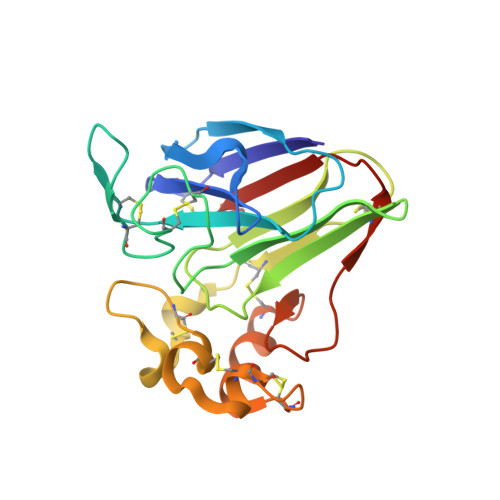
    5FGT, 5FGX

  • PubMed Abstract: 

    Serial femtosecond crystallography (SFX) at X-ray free-electron lasers (XFELs) offers unprecedented possibilities for macromolecular structure determination of systems that are prone to radiation damage. However, phasing XFEL data de novo is complicated by the inherent inaccuracy of SFX data, and only a few successful examples, mostly based on exceedingly strong anomalous or isomorphous difference signals, have been reported. Here, it is shown that SFX data from thaumatin microcrystals can be successfully phased using only the weak anomalous scattering from the endogenous S atoms. Moreover, a step-by-step investigation is presented of the particular problems of SAD phasing of SFX data, analysing data from a derivative with a strong anomalous signal as well as the weak signal from endogenous S atoms.


  • Organizational Affiliation
    • Department of Biomolecular Mechanisms, Max Planck Institute for Medical Research , Jahnstrasse 29, 69120 Heidelberg, Germany.

Macromolecule Content 

  • Total Structure Weight: 22.38 kDa 
  • Atom Count: 1,696 
  • Modeled Residue Count: 207 
  • Deposited Residue Count: 207 
  • Unique protein chains: 1

Macromolecules

Find similar proteins by:|  3D Structure
Entity ID: 1
MoleculeChains  Sequence LengthOrganismDetailsImage
Thaumatin-1207Thaumatococcus danielliiMutation(s): 0 
UniProt
Find proteins for P02883 (Thaumatococcus daniellii)
Explore P02883 
Go to UniProtKB:  P02883
Entity Groups
Sequence Clusters30% Identity50% Identity70% Identity90% Identity95% Identity100% Identity
UniProt GroupP02883
Sequence Annotations
Expand
Reference Sequence

Small Molecules

Ligands 1 Unique
IDChains Name / Formula / InChI Key2D Diagram3D Interactions
TLA

Query on TLA


Download:Ideal Coordinates CCD File
B [auth A]L(+)-TARTARIC ACID
C4 H6 O6
FEWJPZIEWOKRBE-JCYAYHJZSA-N

Experimental Data & Validation

Experimental Data

  • Method: X-RAY DIFFRACTION
  • Resolution: 2.10 Å
  • R-Value Free:  0.186 (Depositor), 0.185 (DCC) 
  • R-Value Work:  0.152 (Depositor), 0.154 (DCC) 
  • R-Value Observed: 0.156 (Depositor) 
Space Group: P 41 21 2
Unit Cell:
Length ( Å )Angle ( ˚ )
a = 58.6α = 90
b = 58.6β = 90
c = 151.3γ = 90
Software Package:
Software NamePurpose
PHENIXrefinement
CrystFELdata reduction
XSCALEdata scaling
autoSHARPphasing

Structure Validation

View Full Validation Report


Entry History 

Revision History  (Full details and data files)

  • Version 1.0: 2016-06-08
    Type: Initial release
  • Version 1.1: 2018-11-14
    Changes: Data collection
  • Version 1.2: 2024-10-23
    Changes: Data collection, Database references, Structure summary