5FBP | pdb_00005fbp

CRYSTAL STRUCTURE OF THE NEUTRAL FORM OF FRUCTOSE-1,6-BISPHOSPHATASE COMPLEXED WITH THE PRODUCT FRUCTOSE 6-PHOSPHATE AT 2.1-ANGSTROMS RESOLUTION


Experimental Data Snapshot

  • Method: X-RAY DIFFRACTION
  • Resolution: 2.10 Å
  • R-Value Work: 
    0.177 (Depositor) 

wwPDB Validation 3D Report Full Report

Validation slider image for 5FBP

This is version 1.5 of the entry. See complete history

Literature

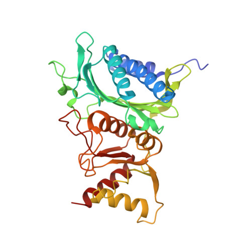
Crystal structure of the neutral form of fructose-1,6-bisphosphatase complexed with the product fructose 6-phosphate at 2.1-A resolution.

Ke, H.M.Zhang, Y.P.Liang, J.Y.Lipscomb, W.N.

(1991) Proc Natl Acad Sci U S A 88: 2989-2993

  • DOI: https://doi.org/10.1073/pnas.88.8.2989
  • Primary Citation Related Structures: 
    5FBP

  • PubMed Abstract: 

    The crystal structure of fructose-1,6-bisphosphatase (EC 3.1.3.11) complexed with the product fructose 6-phosphate (F6P) has been refined at 2.1-A resolution to an R factor of 0.177 with root-mean-square deviations of 0.014 A and 2.9 degrees from the ideal geometries of bond lengths and bond angles, respectively. The secondary structures but not the trace of the unligated enzyme have been slightly revised in the F6P complex at this higher resolution. Helix H4 in the unligated structure has been refined to a helix-like coil, and two very short 3(10) helices have been found, one in H4 and one in H5. F6P at 10 mM concentration in the absence of divalent metals in our study shows major binding at the active site and minor binding at the AMP site. The major site has almost equal full occupancy in the C1 and C2 chains of the crystallographic asymmetric unit, while the minor site shows occupancy only in the C1 chain at about 50%. The electron density in both (2Fo - Fc) and (Fo - Fc) maps calculated by omitting F6P slightly favors the beta anomer of D-F6P over the alpha anomer. Possible functions of the active-site residues are discussed, and candidates are suggested for site-directed mutagenesis.


  • Organizational Affiliation
    • Gibbs Chemical Laboratory, Harvard University, Cambridge, MA 02138.

Macromolecule Content 

  • Total Structure Weight: 73.79 kDa 
  • Atom Count: 5,159 
  • Modeled Residue Count: 629 
  • Deposited Residue Count: 670 
  • Unique protein chains: 1

Macromolecules

Find similar proteins by:|  3D Structure
Entity ID: 1
MoleculeChains  Sequence LengthOrganismDetailsImage
FRUCTOSE 1,6-BISPHOSPHATASE
A, B
335Sus scrofaMutation(s): 0 
EC: 3.1.3.11
UniProt
Find proteins for P00636 (Sus scrofa)
Explore P00636 
Go to UniProtKB:  P00636
Entity Groups
Sequence Clusters30% Identity50% Identity70% Identity90% Identity95% Identity100% Identity
UniProt GroupP00636
Sequence Annotations
Expand
Reference Sequence

Experimental Data & Validation

Experimental Data

  • Method: X-RAY DIFFRACTION
  • Resolution: 2.10 Å
  • R-Value Work:  0.177 (Depositor) 
Space Group: P 32 2 1
Unit Cell:
Length ( Å )Angle ( ˚ )
a = 131.1α = 90
b = 131.1β = 90
c = 69.7γ = 120
Software Package:
Software NamePurpose
X-PLORrefinement

Structure Validation

View Full Validation Report


Entry History 

Deposition Data

Revision History  (Full details and data files)

  • Version 1.0: 1992-07-15
    Type: Initial release
  • Version 1.1: 2008-03-25
    Changes: Version format compliance
  • Version 1.2: 2011-07-13
    Changes: Derived calculations, Version format compliance
  • Version 1.3: 2017-11-29
    Changes: Derived calculations, Other
  • Version 1.4: 2020-07-29
    Type: Remediation
    Reason: Carbohydrate remediation
    Changes: Data collection, Database references, Derived calculations, Structure summary
  • Version 1.5: 2024-03-13
    Changes: Data collection, Database references, Structure summary