5FAH | pdb_00005fah

KALLIKREIN-7 IN COMPLEX WITH COMPOUND1


Experimental Data Snapshot

  • Method: X-RAY DIFFRACTION
  • Resolution: 1.10 Å
  • R-Value Free: 
    0.197 (Depositor), 0.207 (DCC) 
  • R-Value Work: 
    0.188 (Depositor) 
  • R-Value Observed: 
    0.188 (Depositor) 

Starting Model: experimental
View more details

wwPDB Validation 3D Report Full Report

Validation slider image for 5FAH

Ligand Structure Quality Assessment 


This is version 1.4 of the entry. See complete history

Literature

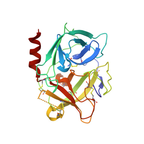
Small-molecule factor D inhibitors targeting the alternative complement pathway.

Maibaum, J.Liao, S.M.Vulpetti, A.Ostermann, N.Randl, S.Rudisser, S.Lorthiois, E.Erbel, P.Kinzel, B.Kolb, F.A.Barbieri, S.Wagner, J.Durand, C.Fettis, K.Dussauge, S.Hughes, N.Delgado, O.Hommel, U.Gould, T.Mac Sweeney, A.Gerhartz, B.Cumin, F.Flohr, S.Schubart, A.Jaffee, B.Harrison, R.Risitano, A.M.Eder, J.Anderson, K.

(2016) Nat Chem Biol 12: 1105-1110

  • DOI: https://doi.org/10.1038/nchembio.2208
  • Primary Citation Related Structures: 
    5FAH, 5FBE, 5FBI, 5FCK, 5FCR

  • PubMed Abstract: 

    Complement is a key component of the innate immune system, recognizing pathogens and promoting their elimination. Complement component 3 (C3) is the central component of the system. Activation of C3 can be initiated by three distinct routes-the classical, the lectin and the alternative pathways-with the alternative pathway also acting as an amplification loop for the other two pathways. The protease factor D (FD) is essential for this amplification process, which, when dysregulated, predisposes individuals to diverse disorders including age-related macular degeneration and paroxysmal nocturnal hemoglobinuria (PNH). Here we describe the identification of potent and selective small-molecule inhibitors of FD. These inhibitors efficiently block alternative pathway (AP) activation and prevent both C3 deposition onto, and lysis of, PNH erythrocytes. Their oral administration inhibited lipopolysaccharide-induced AP activation in FD-humanized mice. These data demonstrate the feasibility of inhibiting the AP with small-molecule antagonists and support the development of FD inhibitors for the treatment of complement-mediated diseases.


  • Organizational Affiliation
    • Novartis Institutes for BioMedical Research, Novartis Pharma AG, Novartis Campus, Basel, Switzerland.

Macromolecule Content 

  • Total Structure Weight: 24.97 kDa 
  • Atom Count: 1,989 
  • Modeled Residue Count: 218 
  • Deposited Residue Count: 224 
  • Unique protein chains: 1

Macromolecules

Find similar proteins by:|  3D Structure
Entity ID: 1
MoleculeChains  Sequence LengthOrganismDetailsImage
Kallikrein-7224Homo sapiensMutation(s): 1 
Gene Names: KLK7PRSS6SCCE
EC: 3.4.21.117
UniProt & NIH Common Fund Data Resources
Find proteins for P49862 (Homo sapiens)
Explore P49862 
Go to UniProtKB:  P49862
PHAROS:  P49862
GTEx:  ENSG00000169035 
Entity Groups
Sequence Clusters30% Identity50% Identity70% Identity90% Identity95% Identity100% Identity
UniProt GroupP49862
Sequence Annotations
Expand
Reference Sequence

Small Molecules

Ligands 2 Unique
IDChains Name / Formula / InChI Key2D Diagram3D Interactions
5VT

Query on 5VT


Download:Ideal Coordinates CCD File
C [auth A](2~{S})-~{N}2-[2-(4-methoxyphenyl)ethyl]-~{N}1-(naphthalen-1-ylmethyl)pyrrolidine-1,2-dicarboxamide
C26 H29 N3 O3
HMNBKWNEJGMBRQ-DEOSSOPVSA-N
ACT

Query on ACT


Download:Ideal Coordinates CCD File
B [auth A]ACETATE ION
C2 H3 O2
QTBSBXVTEAMEQO-UHFFFAOYSA-M
Binding Affinity Annotations 
IDSourceBinding Affinity
5VT BindingDB:  5FAH IC50: 700 (nM) from 1 assay(s)

Experimental Data & Validation

Experimental Data

  • Method: X-RAY DIFFRACTION
  • Resolution: 1.10 Å
  • R-Value Free:  0.197 (Depositor), 0.207 (DCC) 
  • R-Value Work:  0.188 (Depositor) 
  • R-Value Observed: 0.188 (Depositor) 
Space Group: P 21 21 21
Unit Cell:
Length ( Å )Angle ( ˚ )
a = 42.611α = 90
b = 60.343β = 90
c = 80.633γ = 90
Software Package:
Software NamePurpose
DENZOdata reduction
SCALEPACKdata scaling
BUSTERrefinement
MOLREPphasing

Structure Validation

View Full Validation Report


Ligand Structure Quality Assessment 


Entry History 

Deposition Data

Revision History  (Full details and data files)

  • Version 1.0: 2016-10-26
    Type: Initial release
  • Version 1.1: 2016-11-02
    Changes: Database references
  • Version 1.2: 2016-11-23
    Changes: Database references
  • Version 1.3: 2024-01-10
    Changes: Data collection, Database references, Refinement description
  • Version 1.4: 2024-11-13
    Changes: Structure summary