5FA1 | pdb_00005fa1

The structure of the beta-3-deoxy-D-manno-oct-2-ulosonic acid transferase domain of WbbB


Experimental Data Snapshot

  • Method: X-RAY DIFFRACTION
  • Resolution: 2.10 Å
  • R-Value Free: 
    0.210 (Depositor), 0.210 (DCC) 
  • R-Value Work: 
    0.171 (Depositor), 0.172 (DCC) 
  • R-Value Observed: 
    0.173 (Depositor) 

Starting Model: experimental
View more details

wwPDB Validation 3D Report Full Report

Validation slider image for 5FA1

Ligand Structure Quality Assessment 


This is version 1.2 of the entry. See complete history

Literature

Bacterial beta-Kdo glycosyltransferases represent a new glycosyltransferase family (GT99).

Ovchinnikova, O.G.Mallette, E.Koizumi, A.Lowary, T.L.Kimber, M.S.Whitfield, C.

(2016) Proc Natl Acad Sci U S A 113: E3120-E3129

  • DOI: https://doi.org/10.1073/pnas.1603146113
  • Primary Citation Related Structures: 
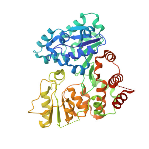
    5FA0, 5FA1

  • PubMed Abstract: 

    Kdo (3-deoxy-d-manno-oct-2-ulosonic acid) is an eight-carbon sugar mostly confined to Gram-negative bacteria. It is often involved in attaching surface polysaccharides to their lipid anchors. α-Kdo provides a bridge between lipid A and the core oligosaccharide in all bacterial LPSs, whereas an oligosaccharide of β-Kdo residues links "group 2" capsular polysaccharides to (lyso)phosphatidylglycerol. β-Kdo is also found in a small number of other bacterial polysaccharides. The structure and function of the prototypical cytidine monophosphate-Kdo-dependent α-Kdo glycosyltransferase from LPS assembly is well characterized. In contrast, the β-Kdo counterparts were not identified as glycosyltransferase enzymes by bioinformatics tools and were not represented among the 98 currently recognized glycosyltransferase families in the Carbohydrate-Active Enzymes database. We report the crystallographic structure and function of a prototype β-Kdo GT from WbbB, a modular protein participating in LPS O-antigen synthesis in Raoultella terrigena The β-Kdo GT has dual Rossmann-fold motifs typical of GT-B enzymes, but extensive deletions, insertions, and rearrangements result in a unique architecture that makes it a prototype for a new GT family (GT99). The cytidine monophosphate-binding site in the C-terminal α/β domain closely resembles the corresponding site in bacterial sialyltransferases, suggesting an evolutionary connection that is not immediately evident from the overall fold or sequence similarities.


  • Organizational Affiliation
    • Department of Molecular and Cellular Biology, University of Guelph, Guelph, ON, Canada N1G 2W1;

Macromolecule Content 

  • Total Structure Weight: 92.84 kDa 
  • Atom Count: 7,100 
  • Modeled Residue Count: 780 
  • Deposited Residue Count: 820 
  • Unique protein chains: 1

Macromolecules

Find similar proteins by:|  3D Structure
Entity ID: 1
MoleculeChains  Sequence LengthOrganismDetailsImage
Putative N-acetyl glucosaminyl transferase
A, B
410Klebsiella terrigenaMutation(s): 0 
Gene Names: wbbB
UniProt
Find proteins for Q6U8B0 (Raoultella terrigena)
Explore Q6U8B0 
Go to UniProtKB:  Q6U8B0
Entity Groups
Sequence Clusters30% Identity50% Identity70% Identity90% Identity95% Identity100% Identity
UniProt GroupQ6U8B0
Sequence Annotations
Expand
Reference Sequence

Experimental Data & Validation

Experimental Data

  • Method: X-RAY DIFFRACTION
  • Resolution: 2.10 Å
  • R-Value Free:  0.210 (Depositor), 0.210 (DCC) 
  • R-Value Work:  0.171 (Depositor), 0.172 (DCC) 
  • R-Value Observed: 0.173 (Depositor) 
Space Group: C 2 2 21
Unit Cell:
Length ( Å )Angle ( ˚ )
a = 92.75α = 90
b = 159.39β = 90
c = 120.04γ = 90
Software Package:
Software NamePurpose
PHENIXrefinement
XDSdata reduction
XSCALEdata scaling
PHASERphasing

Structure Validation

View Full Validation Report


Ligand Structure Quality Assessment 


Entry History 

& Funding Information

Deposition Data


Funding OrganizationLocationGrant Number
GlycoNETCanadaAM-4

Revision History  (Full details and data files)

  • Version 1.0: 2016-05-18
    Type: Initial release
  • Version 1.1: 2018-04-25
    Changes: Data collection, Database references, Derived calculations
  • Version 1.2: 2023-09-27
    Changes: Data collection, Database references, Refinement description