5F9H | pdb_00005f9h

Crystal structure of RIG-I helicase-RD in complex with 24-mer 5' triphosphate hairpin RNA


Experimental Data Snapshot

  • Method: X-RAY DIFFRACTION
  • Resolution: 3.10 Å
  • R-Value Free: 
    0.266 (Depositor), 0.243 (DCC) 
  • R-Value Work: 
    0.213 (Depositor) 
  • R-Value Observed: 
    0.216 (Depositor) 

Starting Model: experimental
View more details

wwPDB Validation 3D Report Full Report

Validation slider image for 5F9H

Ligand Structure Quality Assessment 


This is version 1.4 of the entry. See complete history

Literature

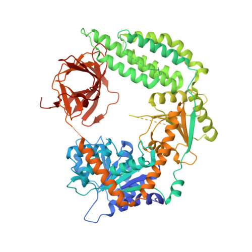
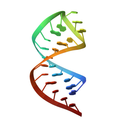
Structural basis for m7G recognition and 2'-O-methyl discrimination in capped RNAs by the innate immune receptor RIG-I.

Devarkar, S.C.Wang, C.Miller, M.T.Ramanathan, A.Jiang, F.Khan, A.G.Patel, S.S.Marcotrigiano, J.

(2016) Proc Natl Acad Sci U S A 113: 596-601

  • DOI: https://doi.org/10.1073/pnas.1515152113
  • Primary Citation Related Structures: 
    5F98, 5F9F, 5F9H

  • PubMed Abstract: 

    RNAs with 5'-triphosphate (ppp) are detected in the cytoplasm principally by the innate immune receptor Retinoic Acid Inducible Gene-I (RIG-I), whose activation triggers a Type I IFN response. It is thought that self RNAs like mRNAs are not recognized by RIG-I because 5'ppp is capped by the addition of a 7-methyl guanosine (m7G) (Cap-0) and a 2'-O-methyl (2'-OMe) group to the 5'-end nucleotide ribose (Cap-1). Here we provide structural and mechanistic basis for exact roles of capping and 2'-O-methylation in evading RIG-I recognition. Surprisingly, Cap-0 and 5'ppp double-stranded (ds) RNAs bind to RIG-I with nearly identical Kd values and activate RIG-I's ATPase and cellular signaling response to similar extents. On the other hand, Cap-0 and 5'ppp single-stranded RNAs did not bind RIG-I and are signaling inactive. Three crystal structures of RIG-I complexes with dsRNAs bearing 5'OH, 5'ppp, and Cap-0 show that RIG-I can accommodate the m7G cap in a cavity created through conformational changes in the helicase-motif IVa without perturbing the ppp interactions. In contrast, Cap-1 modifications abrogate RIG-I signaling through a mechanism involving the H830 residue, which we show is crucial for discriminating between Cap-0 and Cap-1 RNAs. Furthermore, m7G capping works synergistically with 2'-O-methylation to weaken RNA affinity by 200-fold and lower ATPase activity. Interestingly, a single H830A mutation restores both high-affinity binding and signaling activity with 2'-O-methylated dsRNAs. Our work provides new structural insights into the mechanisms of host and viral immune evasion from RIG-I, explaining the complexity of cap structures over evolution.


  • Organizational Affiliation
    • Department of Biochemistry and Molecular Biology, Robert Wood Johnson Medical School, Rutgers University, Piscataway, NJ 08854;

Macromolecule Content 

  • Total Structure Weight: 525.12 kDa 
  • Atom Count: 33,602 
  • Modeled Residue Count: 4,003 
  • Deposited Residue Count: 4,308 
  • Unique protein chains: 1
  • Unique nucleic acid chains: 1

Macromolecules


Find similar proteins by:|  3D Structure
Entity ID: 1
MoleculeChains  Sequence LengthOrganismDetailsImage
Probable ATP-dependent RNA helicase DDX58
A, C, E, G, I
A, C, E, G, I, K
695Homo sapiensMutation(s): 0 
Gene Names: DDX58
EC: 3.6.4.13
UniProt & NIH Common Fund Data Resources
Find proteins for O95786 (Homo sapiens)
Explore O95786 
Go to UniProtKB:  O95786
PHAROS:  O95786
GTEx:  ENSG00000107201 
Entity Groups
Sequence Clusters30% Identity50% Identity70% Identity90% Identity95% Identity100% Identity
UniProt GroupO95786
Sequence Annotations
Expand
Reference Sequence
Find similar nucleic acids by:  Sequence
Entity ID: 2
MoleculeChains LengthOrganismImage
RNA (5'-R(P*AP*AP*UP*AP*UP*AP*AP*UP*AP*GP*UP*GP*AP*UP*AP*UP*UP*AP*UP*AP*UP*UP*C)-3')
B, D, F, H, J
B, D, F, H, J, L
23synthetic construct
Sequence Annotations
Expand
Reference Sequence

Small Molecules

Ligands 3 Unique
IDChains Name / Formula / InChI Key2D Diagram3D Interactions
GTP

Query on GTP


Download:Ideal Coordinates CCD File
CA [auth J]
FA [auth L]
Q [auth B]
S [auth D]
V [auth F]
CA [auth J],
FA [auth L],
Q [auth B],
S [auth D],
V [auth F],
Z [auth H]
GUANOSINE-5'-TRIPHOSPHATE
C10 H16 N5 O14 P3
XKMLYUALXHKNFT-UUOKFMHZSA-N
ZN

Query on ZN


Download:Ideal Coordinates CCD File
AA [auth I]
DA [auth K]
M [auth A]
R [auth C]
T [auth E]
AA [auth I],
DA [auth K],
M [auth A],
R [auth C],
T [auth E],
W [auth G]
ZINC ION
Zn
PTFCDOFLOPIGGS-UHFFFAOYSA-N
MG

Query on MG


Download:Ideal Coordinates CCD File
BA [auth I]
EA [auth K]
N [auth A]
O [auth A]
P [auth A]
BA [auth I],
EA [auth K],
N [auth A],
O [auth A],
P [auth A],
U [auth E],
X [auth G],
Y [auth G]
MAGNESIUM ION
Mg
JLVVSXFLKOJNIY-UHFFFAOYSA-N

Experimental Data & Validation

Experimental Data

  • Method: X-RAY DIFFRACTION
  • Resolution: 3.10 Å
  • R-Value Free:  0.266 (Depositor), 0.243 (DCC) 
  • R-Value Work:  0.213 (Depositor) 
  • R-Value Observed: 0.216 (Depositor) 
Space Group: P 21 21 21
Unit Cell:
Length ( Å )Angle ( ˚ )
a = 111.18α = 90
b = 174.81β = 90
c = 309.28γ = 90
Software Package:
Software NamePurpose
PHENIXrefinement
iMOSFLMdata reduction
Aimlessdata scaling
PHENIXphasing

Structure Validation

View Full Validation Report


Ligand Structure Quality Assessment 


Entry History 

Deposition Data

Revision History  (Full details and data files)

  • Version 1.0: 2016-01-13
    Type: Initial release
  • Version 1.1: 2016-01-20
    Changes: Database references, Structure summary
  • Version 1.2: 2016-01-27
    Changes: Database references
  • Version 1.3: 2016-04-06
    Changes: Other
  • Version 1.4: 2023-09-27
    Changes: Data collection, Database references, Derived calculations, Refinement description