5F6B

Crystal structure of the Q108K:K40L:T51V:R58Y:Y19W mutant of human Cellular Retinol Binding Protein II in complex with All-trans-Retinal at 1.3 Angstrom Resolution


Experimental Data Snapshot

  • Method: X-RAY DIFFRACTION
  • Resolution: 1.31 Å
  • R-Value Free: 
    0.235 (Depositor), 0.230 (DCC) 
  • R-Value Work: 
    0.201 (Depositor), 0.200 (DCC) 
  • R-Value Observed: 
    0.203 (Depositor) 

Starting Model: experimental
View more details

wwPDB Validation   3D Report Full Report


Ligand Structure Quality Assessment 

Created with Raphaël 2.3.0Worse 01 BetterLigand structure goodness of fit to experimental dataBest fitted RETClick on this verticalbar to view details

This is version 1.2 of the entry. See complete history


Literature

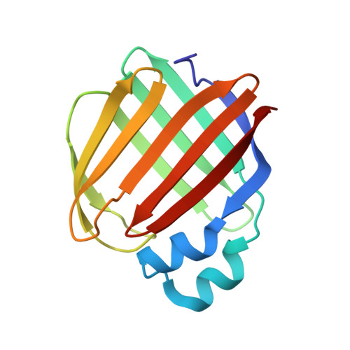
Crystal structure of the Q108K:K40L:T51V:R58Y:Y19W mutant of human Cellular Retinol Binding ProteinII in complex with All-trans-Retinal at 1.31 Angstrom Resolution

Nossoni, Z.Nosrati, M.Wang, W.Berbasova, T.Vasileiou, C.Borhan, B.Geiger, J.H.

To be published.

Macromolecules
Find similar proteins by:  (by identity cutoff)  |  3D Structure
Entity ID: 1
MoleculeChains Sequence LengthOrganismDetailsImage
Retinol-binding protein 2
A, B
133Homo sapiensMutation(s): 5 
Gene Names: RBP2CRBP2
UniProt & NIH Common Fund Data Resources
Find proteins for P50120 (Homo sapiens)
Explore P50120 
Go to UniProtKB:  P50120
PHAROS:  P50120
GTEx:  ENSG00000114113 
Entity Groups  
Sequence Clusters30% Identity50% Identity70% Identity90% Identity95% Identity100% Identity
UniProt GroupP50120
Sequence Annotations
Expand
  • Reference Sequence
Experimental Data & Validation

Experimental Data

  • Method: X-RAY DIFFRACTION
  • Resolution: 1.31 Å
  • R-Value Free:  0.235 (Depositor), 0.230 (DCC) 
  • R-Value Work:  0.201 (Depositor), 0.200 (DCC) 
  • R-Value Observed: 0.203 (Depositor) 
Space Group: P 1
Unit Cell:
Length ( Å )Angle ( ˚ )
a = 29.953α = 90.85
b = 35.721β = 88.45
c = 64.937γ = 65.68
Software Package:
Software NamePurpose
PHENIXrefinement
HKL-2000data reduction
HKL-2000data scaling
MOLREPphasing

Structure Validation

View Full Validation Report


Ligand Structure Quality Assessment 

Created with Raphaël 2.3.0Worse 01 BetterLigand structure goodness of fit to experimental dataBest fitted RETClick on this verticalbar to view details

Entry History 

Deposition Data

Revision History  (Full details and data files)

  • Version 1.0: 2016-12-07
    Type: Initial release
  • Version 1.1: 2023-09-27
    Changes: Data collection, Database references, Refinement description
  • Version 1.2: 2024-10-30
    Changes: Structure summary