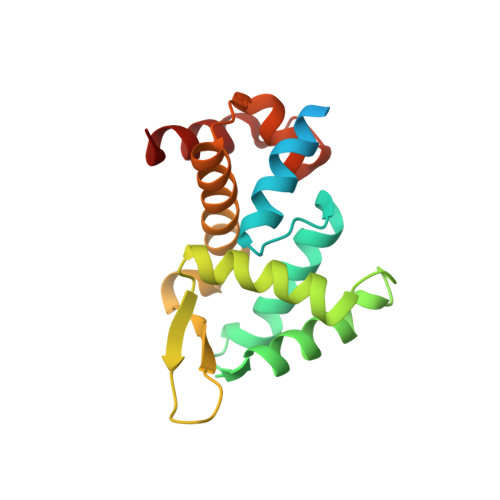
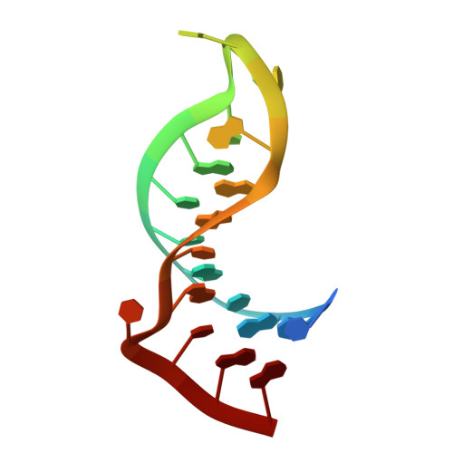
5F5H | pdb_00005f5h

X-ray structure of Roquin ROQ domain in complex with Ox40 hexa-loop RNA motif


Experimental Data Snapshot

  • Method: X-RAY DIFFRACTION
  • Resolution: 2.23 Å
  • R-Value Free: 
    0.257 (Depositor), 0.259 (DCC) 
  • R-Value Work: 
    0.216 (Depositor), 0.220 (DCC) 
  • R-Value Observed: 
    0.218 (Depositor) 

Starting Model: experimental
View more details

wwPDB Validation 3D Report Full Report

Validation slider image for 5F5H

This is version 1.1 of the entry. See complete history. 

Literature

Roquin recognizes a non-canonical hexaloop structure in the 3'-UTR of Ox40.

Janowski, R., Heinz, G.A., Schlundt, A., Wommelsdorf, N., Brenner, S., Gruber, A.R., Blank, M., Buch, T., Buhmann, R., Zavolan, M., Niessing, D., Heissmeyer, V., Sattler, M.

(2016) Nat Commun 7: 11032-11032

  • DOI: https://doi.org/10.1038/ncomms11032
  • Primary Citation Related Structures: 
    5F5F, 5F5H

  • PubMed Abstract: 

    The RNA-binding protein Roquin is required to prevent autoimmunity. Roquin controls T-helper cell activation and differentiation by limiting the induced expression of costimulatory receptors such as tumor necrosis factor receptor superfamily 4 (Tnfrs4 or Ox40). A constitutive decay element (CDE) with a characteristic triloop hairpin was previously shown to be recognized by Roquin. Here we use SELEX assays to identify a novel U-rich hexaloop motif, representing an alternative decay element (ADE). Crystal structures and NMR data show that the Roquin-1 ROQ domain recognizes hexaloops in the SELEX-derived ADE and in an ADE-like variant present in the Ox40 3'-UTR with identical binding modes. In cells, ADE-like and CDE-like motifs cooperate in the repression of Ox40 by Roquin. Our data reveal an unexpected recognition of hexaloop cis elements for the posttranscriptional regulation of target messenger RNAs by Roquin.


  • Organizational Affiliation: 
    • Group Intracellular Transport and RNA Biology, Institute of Structural Biology, Helmholtz Zentrum München, Ingolstädter Landstrasse 1, Neuherberg DE-85764, Germany.

Macromolecule Content 

  • Total Structure Weight: 55.12 kDa 
  • Atom Count: 3,397 
  • Modeled Residue Count: 342 
  • Deposited Residue Count: 404 
  • Unique protein chains: 1
  • Unique nucleic acid chains: 1

Macromolecules


Find similar proteins by:|  3D Structure
Entity ID: 1
MoleculeChains  Sequence LengthOrganismDetailsImage
Roquin-1
A, B
180Mus musculusMutation(s): 0 
Gene Names: Rc3h1, Gm551, Kiaa2025
EC: 2.3.2.27
UniProt
Find proteins for Q4VGL6 (Mus musculus)
Explore Q4VGL6 
Go to UniProtKB:  Q4VGL6
Entity Groups
Sequence Clusters30% Identity50% Identity70% Identity90% Identity95% Identity100% Identity
UniProt GroupQ4VGL6
Sequence Annotations
Expand
Reference Sequence
Find similar nucleic acids by:  Sequence
Entity ID: 2
MoleculeChains LengthOrganismImage
RNA (5'-R(P*CP*CP*AP*CP*AP*CP*CP*GP*UP*UP*CP*UP*AP*GP*GP*UP*GP*CP*UP*GP*G)-3')
C, D
22Mus musculus
Sequence Annotations
Expand
Reference Sequence

Small Molecules

Ligands 1 Unique
IDChains Name / Formula / InChI Key2D Diagram3D Interactions
GOL

Query on GOL


Download:Ideal Coordinates CCD File
E [auth A]GLYCEROL
C3 H8 O3
PEDCQBHIVMGVHV-UHFFFAOYSA-N

Experimental Data & Validation

Experimental Data

  • Method: X-RAY DIFFRACTION
  • Resolution: 2.23 Å
  • R-Value Free:  0.257 (Depositor), 0.259 (DCC) 
  • R-Value Work:  0.216 (Depositor), 0.220 (DCC) 
  • R-Value Observed: 0.218 (Depositor) 
Space Group: P 21 21 2
Unit Cell:
Length ( Å )Angle ( ˚ )
a = 89.66α = 90
b = 115.79β = 90
c = 42.61γ = 90
Software Package:
Software NamePurpose
REFMACrefinement
XDSdata reduction
XSCALEdata scaling
PHASERphasing

Structure Validation

View Full Validation Report


Entry History 

Deposition Data

Revision History  (Full details and data files)

  • Version 1.0: 2016-04-06
    Type: Initial release
  • Version 1.1: 2024-01-10
    Changes: Data collection, Database references, Refinement description