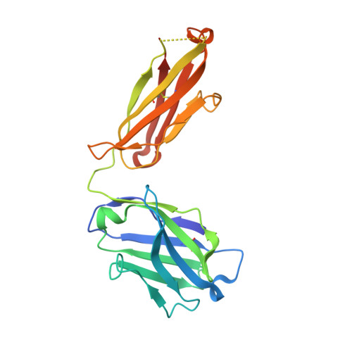
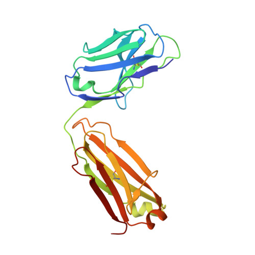
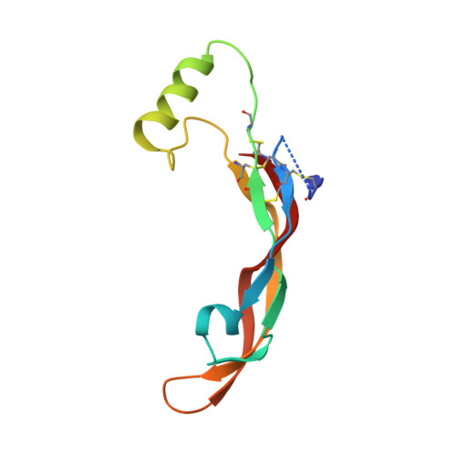
5F3H | pdb_00005f3h

Structure of myostatin in complex with humanized RK35 antibody


Experimental Data Snapshot

  • Method: X-RAY DIFFRACTION
  • Resolution: 2.70 Å
  • R-Value Free: 
    0.270 (Depositor), 0.238 (DCC) 
  • R-Value Work: 
    0.218 (Depositor) 
  • R-Value Observed: 
    0.221 (Depositor) 

Starting Model: experimental
View more details

wwPDB Validation 3D Report Full Report

Validation slider image for 5F3H

This is version 1.3 of the entry. See complete history. 

Literature

Beyond CDR-grafting: Structure-guided humanization of framework and CDR regions of an anti-myostatin antibody.

Apgar, J.R., Mader, M., Agostinelli, R., Benard, S., Bialek, P., Johnson, M., Gao, Y., Krebs, M., Owens, J., Parris, K., St Andre, M., Svenson, K., Morris, C., Tchistiakova, L.

(2016) MAbs 8: 1302-1318

  • DOI: https://doi.org/10.1080/19420862.2016.1215786
  • Primary Citation Related Structures: 
    5F3B, 5F3H

  • PubMed Abstract: 

    Antibodies are an important class of biotherapeutics that offer specificity to their antigen, long half-life, effector function interaction and good manufacturability. The immunogenicity of non-human-derived antibodies, which can be a major limitation to development, has been partially overcome by humanization through complementarity-determining region (CDR) grafting onto human acceptor frameworks. The retention of foreign content in the CDR regions, however, is still a potential immunogenic liability. Here, we describe the humanization of an anti-myostatin antibody utilizing a 2-step process of traditional CDR-grafting onto a human acceptor framework, followed by a structure-guided approach to further reduce the murine content of CDR-grafted antibodies. To accomplish this, we solved the co-crystal structures of myostatin with the chimeric (Protein Databank (PDB) id 5F3B) and CDR-grafted anti-myostatin antibody (PDB id 5F3H), allowing us to computationally predict the structurally important CDR residues as well as those making significant contacts with the antigen. Structure-based rational design enabled further germlining of the CDR-grafted antibody, reducing the murine content of the antibody without affecting antigen binding. The overall "humanness" was increased for both the light and heavy chain variable regions.


  • Organizational Affiliation: 
    • a Biomedicine Design, Pfizer Inc. , Cambridge , MA , USA.

Macromolecule Content 

  • Total Structure Weight: 236.2 kDa 
  • Atom Count: 15,926 
  • Modeled Residue Count: 2,099 
  • Deposited Residue Count: 2,172 
  • Unique protein chains: 3

Macromolecules

Find similar proteins by:|  3D Structure
Entity ID: 1
MoleculeChains  Sequence LengthOrganismDetailsImage
humanized RK35 antibody heavy chain
A, C, E, G
221Mus musculusMutation(s): 0 
Entity Groups
Sequence Clusters30% Identity50% Identity70% Identity90% Identity95% Identity100% Identity
Sequence Annotations
Expand
Reference Sequence
Find similar proteins by:|  3D Structure
Entity ID: 2
MoleculeChains  Sequence LengthOrganismDetailsImage
humanized RK35 antibody light chain
B, D, F, H
214Mus musculusMutation(s): 0 
Entity Groups
Sequence Clusters30% Identity50% Identity70% Identity90% Identity95% Identity100% Identity
Sequence Annotations
Expand
Reference Sequence
Find similar proteins by:|  3D Structure
Entity ID: 3
MoleculeChains  Sequence LengthOrganismDetailsImage
Growth/differentiation factor 8
I, J, K, L
108Homo sapiensMutation(s): 0 
Gene Names: MSTN, GDF8
UniProt & NIH Common Fund Data Resources
Find proteins for O14793 (Homo sapiens)
Explore O14793 
Go to UniProtKB:  O14793
PHAROS:  O14793
GTEx:  ENSG00000138379 
Entity Groups
Sequence Clusters30% Identity50% Identity70% Identity90% Identity95% Identity100% Identity
UniProt GroupO14793
Sequence Annotations
Expand
Reference Sequence

Experimental Data & Validation

Experimental Data

  • Method: X-RAY DIFFRACTION
  • Resolution: 2.70 Å
  • R-Value Free:  0.270 (Depositor), 0.238 (DCC) 
  • R-Value Work:  0.218 (Depositor) 
  • R-Value Observed: 0.221 (Depositor) 
Space Group: P 1
Unit Cell:
Length ( Å )Angle ( ˚ )
a = 75.138α = 88.33
b = 80.916β = 103.78
c = 101.401γ = 92.4
Software Package:
Software NamePurpose
PHENIXrefinement
SCALEPACKdata scaling
PDB_EXTRACTdata extraction
HKL-2000data reduction
AMoREphasing

Structure Validation

View Full Validation Report


Entry History 

Deposition Data

Revision History  (Full details and data files)

  • Version 1.0: 2016-09-28
    Type: Initial release
  • Version 1.1: 2016-10-12
    Changes: Database references
  • Version 1.2: 2023-09-27
    Changes: Data collection, Database references, Derived calculations, Refinement description
  • Version 1.3: 2024-10-23
    Changes: Structure summary