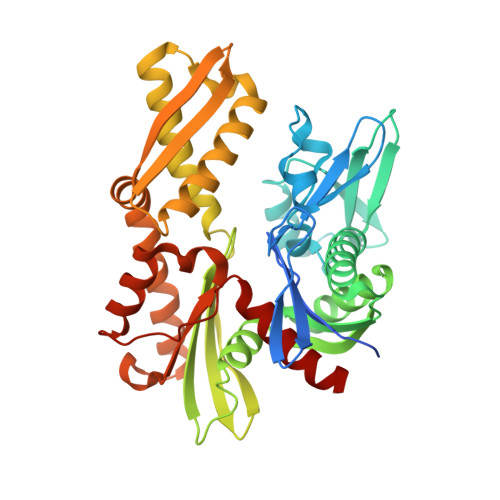
Probing the ATP Site of GRP78 with Nucleotide Triphosphate Analogs.
Hughes, S.J., Antoshchenko, T., Chen, Y., Lu, H., Pizarro, J.C., Park, H.W.(2016) PLoS One 11: e0154862-e0154862
- PubMed: 27144892
- DOI: https://doi.org/10.1371/journal.pone.0154862
- Primary Citation of Related Structures:
5EVZ, 5EX5, 5EXW, 5EY4, 5F0X, 5F1X, 5F2R - PubMed Abstract:
GRP78, a member of the ER stress protein family, can relocate to the surface of cancer cells, playing key roles in promoting cell proliferation and metastasis. GRP78 consists of two major functional domains: the ATPase and protein/peptide-binding domains. The protein/peptide-binding domain of cell-surface GRP78 has served as a novel functional receptor for delivering cytotoxic agents (e.g., a apoptosis-inducing peptide or taxol) across the cell membrane. Here, we report our study on the ATPase domain of GRP78 (GRP78ATPase), whose potential as a transmembrane delivery system of cytotoxic agents (e.g., ATP-based nucleotide triphosphate analogs) remains unexploited. As the binding of ligands (ATP analogs) to a receptor (GRP78ATPase) is a pre-requisite for internalization, we determined the binding affinities and modes of GRP78ATPase for ADP, ATP and several ATP analogs using surface plasmon resonance and x-ray crystallography. The tested ATP analogs contain one of the following modifications: the nitrogen at the adenine ring 7-position to a carbon atom (7-deazaATP), the oxygen at the β-γ bridge position to a carbon atom (AMPPCP), or the removal of the 2'-OH group (2'-deoxyATP). We found that 7-deazaATP displays an affinity and a binding mode that resemble those of ATP regardless of magnesium ion (Mg++) concentration, suggesting that GRP78 is tolerant to modifications at the 7-position. By comparison, AMPPCP's binding affinity was lower than ATP and Mg++-dependent, as the removal of Mg++ nearly abolished binding to GRP78ATPase. The AMPPCP-Mg++ structure showed evidence for the critical role of Mg++ in AMPPCP binding affinity, suggesting that while GRP78 is sensitive to modifications at the β-γ bridge position, these can be tolerated in the presence of Mg++. Furthermore, 2'-deoxyATP's binding affinity was significantly lower than those for all other nucleotides tested, even in the presence of Mg++. The 2'-deoxyATP structure showed the conformation of the bound nucleotide flipped out of the active site, explaining the low affinity binding to GRP78 and suggesting that the 2'-OH group is essential for the high affinity binding to GRP78. Together, our results demonstrate that GRP78ATPase possesses nucleotide specificity more relaxed than previously anticipated and can tolerate certain modifications to the nucleobase 7-position and, to a lesser extent, the β-γ bridging atom, thereby providing a possible atomic mechanism underlying the transmembrane transport of the ATP analogs.
Organizational Affiliation:
Department of Pharmacology and Toxicology, University of Toronto, Toronto, Ontario M5G 1L7, Canada.


