5F0V | pdb_00005f0v

X-ray crystal structure of a thiolase from Escherichia coli at 1.8 A resolution


Experimental Data Snapshot

  • Method: X-RAY DIFFRACTION
  • Resolution: 1.80 Å
  • R-Value Free: 
    0.195 (Depositor), 0.206 (DCC) 
  • R-Value Work: 
    0.160 (Depositor), 0.172 (DCC) 
  • R-Value Observed: 
    0.162 (Depositor) 

Starting Model: experimental
View more details

wwPDB Validation 3D Report Full Report

Validation slider image for 5F0V

This is version 1.2 of the entry. See complete history

Literature

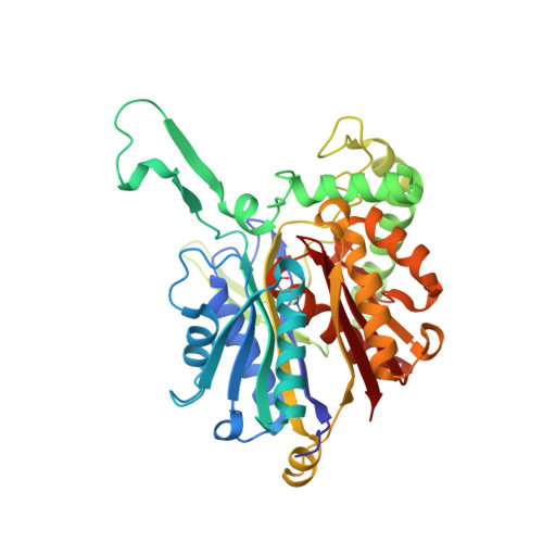
Crystal structure of a thiolase from Escherichia coli at 1.8 angstrom resolution.

Ithayaraja, M.Janardan, N.Wierenga, R.K.Savithri, H.S.Murthy, M.R.

(2016) Acta Crystallogr F Struct Biol Commun 72: 534-544

  • DOI: https://doi.org/10.1107/S2053230X16008451
  • Primary Citation Related Structures: 
    5F0V, 5F38

  • PubMed Abstract: 

    Thiolases catalyze the Claisen condensation of two acetyl-CoA molecules to give acetoacetyl-CoA, as well as the reverse degradative reaction. Four genes coding for thiolases or thiolase-like proteins are found in the Escherichia coli genome. In this communication, the successful cloning, purification, crystallization and structure determination at 1.8 Å resolution of a homotetrameric E. coli thiolase are reported. The structure of E. coli thiolase co-crystallized with acetyl-CoA at 1.9 Å resolution is also reported. As observed in other tetrameric thiolases, the present E. coli thiolase is a dimer of two tight dimers and probably functions as a biodegradative enzyme. Comparison of the structure and biochemical properties of the E. coli enzyme with those of other well studied thiolases reveals certain novel features of this enzyme, such as the modification of a lysine in the dimeric interface, the possible oxidation of the catalytic Cys88 in the structure of the enzyme obtained in the presence of CoA and active-site hydration. The tetrameric enzyme also displays an interesting departure from exact 222 symmetry, which is probably related to the deformation of the tetramerization domain that stabilizes the oligomeric structure of the protein. The current study allows the identification of substrate-binding amino-acid residues and water networks at the active site and provides the structural framework required for understanding the biochemical properties as well as the physiological function of this E. coli thiolase.


  • Organizational Affiliation
    • Molecular Biophysics Unit, Indian Institute of Science, Bangalore, Karnataka 560 012, India.

Macromolecule Content 

  • Total Structure Weight: 164.58 kDa 
  • Atom Count: 12,462 
  • Modeled Residue Count: 1,579 
  • Deposited Residue Count: 1,580 
  • Unique protein chains: 1

Macromolecules

Find similar proteins by:|  3D Structure
Entity ID: 1
MoleculeChains  Sequence LengthOrganismDetailsImage
Acetyl-CoA acetyltransferase
A, B, C, D
395Escherichia coli K-12Mutation(s): 0 
Gene Names: atoBb2224JW2218
EC: 2.3.1.9
UniProt
Find proteins for P76461 (Escherichia coli (strain K12))
Explore P76461 
Go to UniProtKB:  P76461
Entity Groups
Sequence Clusters30% Identity50% Identity70% Identity90% Identity95% Identity100% Identity
UniProt GroupP76461
Sequence Annotations
Expand
Reference Sequence

Small Molecules

Ligands 1 Unique
IDChains Name / Formula / InChI Key2D Diagram3D Interactions
EDO

Query on EDO


Download:Ideal Coordinates CCD File
AA [auth B]
BA [auth B]
CA [auth B]
DA [auth B]
E [auth A]
AA [auth B],
BA [auth B],
CA [auth B],
DA [auth B],
E [auth A],
EA [auth B],
F [auth A],
FA [auth B],
G [auth A],
GA [auth B],
H [auth A],
HA [auth B],
I [auth A],
IA [auth C],
J [auth A],
JA [auth C],
K [auth A],
KA [auth C],
L [auth A],
LA [auth C],
M [auth A],
MA [auth C],
N [auth A],
NA [auth C],
O [auth A],
OA [auth D],
P [auth A],
PA [auth D],
Q [auth A],
QA [auth D],
R [auth A],
RA [auth D],
S [auth A],
SA [auth D],
T [auth A],
TA [auth D],
U [auth B],
UA [auth D],
V [auth B],
VA [auth D],
W [auth B],
X [auth B],
Y [auth B],
Z [auth B]
1,2-ETHANEDIOL
C2 H6 O2
LYCAIKOWRPUZTN-UHFFFAOYSA-N
Modified Residues  1 Unique
IDChains TypeFormula2D DiagramParent
MLY
Query on MLY
A, B, C, D
L-PEPTIDE LINKINGC8 H18 N2 O2LYS

Experimental Data & Validation

Experimental Data

  • Method: X-RAY DIFFRACTION
  • Resolution: 1.80 Å
  • R-Value Free:  0.195 (Depositor), 0.206 (DCC) 
  • R-Value Work:  0.160 (Depositor), 0.172 (DCC) 
  • R-Value Observed: 0.162 (Depositor) 
Space Group: C 1 2 1
Unit Cell:
Length ( Å )Angle ( ˚ )
a = 190.93α = 90
b = 75.31β = 131.42
c = 147.41γ = 90
Software Package:
Software NamePurpose
REFMACrefinement
SCALAdata reduction
SCALAdata scaling
PHASERphasing

Structure Validation

View Full Validation Report


Entry History 

& Funding Information

Deposition Data


Funding OrganizationLocationGrant Number
DBTIndiaBT/IN/FINNISH/27/MRNM/2009

Revision History  (Full details and data files)

  • Version 1.0: 2016-07-13
    Type: Initial release
  • Version 1.1: 2019-12-18
    Changes: Database references, Derived calculations
  • Version 1.2: 2023-11-08
    Changes: Data collection, Database references, Refinement description