5EZO | pdb_00005ezo

Crystal Structure of PfCyRPA in complex with an invasion-inhibitory antibody Fab.


Experimental Data Snapshot

  • Method: X-RAY DIFFRACTION
  • Resolution: 3.63 Å
  • R-Value Free: 
    0.330 (Depositor), 0.323 (DCC) 
  • R-Value Work: 
    0.274 (Depositor), 0.282 (DCC) 
  • R-Value Observed: 
    0.276 (Depositor) 

Starting Models: experimental
View more details

wwPDB Validation 3D Report Full Report

Validation slider image for 5EZO

This is version 2.2 of the entry. See complete history

Literature

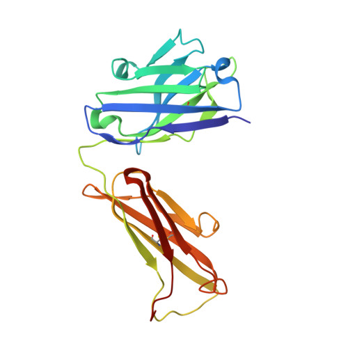
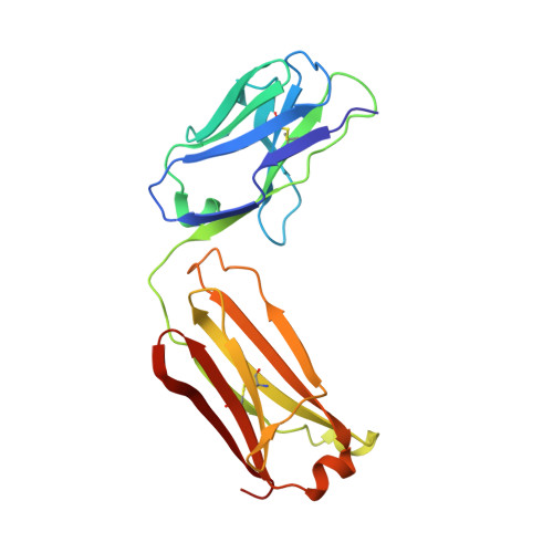
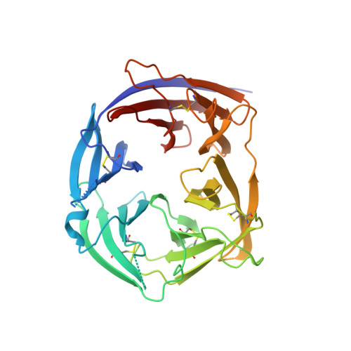
Structure of the malaria vaccine candidate antigen CyRPA and its complex with a parasite invasion inhibitory antibody.

Favuzza, P.Guffart, E.Tamborrini, M.Scherer, B.Dreyer, A.M.Rufer, A.C.Erny, J.Hoernschemeyer, J.Thoma, R.Schmid, G.Gsell, B.Lamelas, A.Benz, J.Joseph, C.Matile, H.Pluschke, G.Rudolph, M.G.

(2017) Elife 6

  • DOI: https://doi.org/10.7554/eLife.20383
  • Primary Citation Related Structures: 
    5EZI, 5EZJ, 5EZL, 5EZN, 5EZO

  • PubMed Abstract: 

    Invasion of erythrocytes by Plasmodial merozoites is a composite process involving the interplay of several proteins. Among them, the Plasmodium falciparum Cysteine-Rich Protective Antigen (PfCyRPA) is a crucial component of a ternary complex, including Reticulocyte binding-like Homologous protein 5 (PfRH5) and the RH5-interacting protein (PfRipr), essential for erythrocyte invasion. Here, we present the crystal structures of PfCyRPA and its complex with the antigen-binding fragment of a parasite growth inhibitory antibody. PfCyRPA adopts a 6-bladed β-propeller structure with similarity to the classic sialidase fold, but it has no sialidase activity and fulfills a purely non-enzymatic function. Characterization of the epitope recognized by protective antibodies may facilitate design of peptidomimetics to focus vaccine responses on protective epitopes. Both in vitro and in vivo anti-PfCyRPA and anti-PfRH5 antibodies showed more potent parasite growth inhibitory activity in combination than on their own, supporting a combined delivery of PfCyRPA and PfRH5 in vaccines.


  • Organizational Affiliation
    • Medical Parasitology and Infection Biology Department, Swiss Tropical and Public Health Institute, Basel, Switzerland.

Macromolecule Content 

  • Total Structure Weight: 87.14 kDa 
  • Atom Count: 6,033 
  • Modeled Residue Count: 756 
  • Deposited Residue Count: 767 
  • Unique protein chains: 3

Macromolecules

Find similar proteins by:|  3D Structure
Entity ID: 1
MoleculeChains  Sequence LengthOrganismDetailsImage
PfCyRPAA [auth H]222Homo sapiensMutation(s): 0 
Find similar proteins by:|  3D Structure
Entity ID: 2
MoleculeChains  Sequence LengthOrganismDetailsImage
c12 FABB [auth L]213Mus musculusMutation(s): 0 
Find similar proteins by:|  3D Structure
Entity ID: 3
MoleculeChains  Sequence LengthOrganismDetailsImage
c12 FabC [auth A]332Plasmodium falciparum 3D7Mutation(s): 0 
Gene Names: PFD1130w
UniProt
Find proteins for Q8IFM8 (Plasmodium falciparum (isolate 3D7))
Explore Q8IFM8 
Go to UniProtKB:  Q8IFM8
Entity Groups
Sequence Clusters30% Identity50% Identity70% Identity90% Identity95% Identity100% Identity
UniProt GroupQ8IFM8
Sequence Annotations
Expand
Reference Sequence

Oligosaccharides

Help  
Entity ID: 4
MoleculeChains Length2D Diagram GlycosylationD Interactions
2-acetamido-2-deoxy-beta-D-glucopyranose-(1-4)-[alpha-L-fucopyranose-(1-6)]2-acetamido-2-deoxy-beta-D-glucopyranoseD [auth B]3N-Glycosylation
Glycosylation Resources
GlyTouCan: G21290RB
GlyCosmos: G21290RB
GlyGen: G21290RB

Experimental Data & Validation

Experimental Data

  • Method: X-RAY DIFFRACTION
  • Resolution: 3.63 Å
  • R-Value Free:  0.330 (Depositor), 0.323 (DCC) 
  • R-Value Work:  0.274 (Depositor), 0.282 (DCC) 
  • R-Value Observed: 0.276 (Depositor) 
Space Group: P 1 21 1
Unit Cell:
Length ( Å )Angle ( ˚ )
a = 95.801α = 90
b = 44.681β = 108.03
c = 121.336γ = 90
Software Package:
Software NamePurpose
XSCALEdata scaling
PDB_EXTRACTdata extraction
PHENIXrefinement
XDSdata reduction
PHASERphasing

Structure Validation

View Full Validation Report


Entry History 

Deposition Data

Revision History  (Full details and data files)

  • Version 1.0: 2017-01-11
    Type: Initial release
  • Version 1.1: 2017-02-15
    Changes: Structure summary
  • Version 1.2: 2017-03-01
    Changes: Database references
  • Version 1.3: 2017-10-11
    Changes: Data collection
  • Version 2.0: 2020-07-29
    Type: Remediation
    Reason: Carbohydrate remediation
    Changes: Atomic model, Data collection, Derived calculations, Structure summary
  • Version 2.1: 2024-01-10
    Changes: Data collection, Database references, Derived calculations, Refinement description, Structure summary
  • Version 2.2: 2024-11-20
    Changes: Structure summary