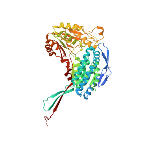
1.72 Angstrom resolution crystal structure of betaine aldehyde dehydrogenase (betB) P449M point mutant from Staphylococcus aureus in complex with NAD+ and BME-modified Cys289
Halavaty, A.S., Minasov, G., Chen, C., Joo, J.C., Yakunin, A.F., Anderson, W.F.To be published.