5EYA | pdb_00005eya

TRIM25 RING domain in complex with Ubc13-Ub conjugate


Experimental Data Snapshot

  • Method: X-RAY DIFFRACTION
  • Resolution: 2.40 Å
  • R-Value Free: 
    0.228 (Depositor), 0.230 (DCC) 
  • R-Value Work: 
    0.189 (Depositor), 0.195 (DCC) 
  • R-Value Observed: 
    0.192 (Depositor) 

wwPDB Validation 3D Report Full Report

Validation slider image for 5EYA

This is version 1.3 of the entry. See complete history

Literature

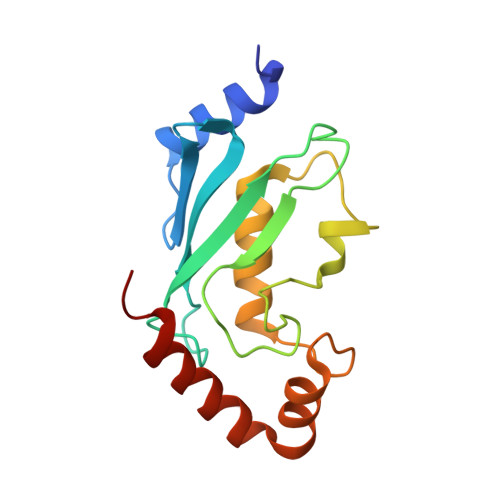
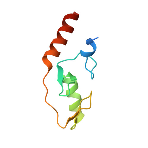
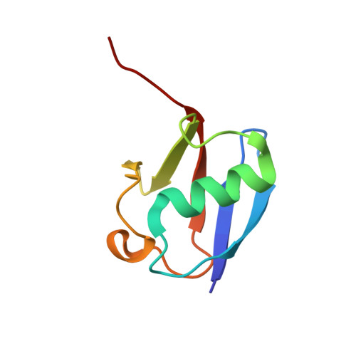
Mechanism of TRIM25 Catalytic Activation in the Antiviral RIG-I Pathway.

Sanchez, J.G.Chiang, J.J.Sparrer, K.M.Alam, S.L.Chi, M.Roganowicz, M.D.Sankaran, B.Gack, M.U.Pornillos, O.

(2016) Cell Rep 16: 1315-1325

  • DOI: https://doi.org/10.1016/j.celrep.2016.06.070
  • Primary Citation Related Structures: 
    5EYA

  • PubMed Abstract: 

    Antiviral response pathways induce interferon by higher-order assembly of signaling complexes called signalosomes. Assembly of the RIG-I signalosome is regulated by K63-linked polyubiquitin chains, which are synthesized by the E3 ubiquitin ligase, TRIM25. We have previously shown that the TRIM25 coiled-coil domain is a stable, antiparallel dimer that positions two catalytic RING domains on opposite ends of an elongated rod. We now show that the RING domain is a separate self-association motif that engages ubiquitin-conjugated E2 enzymes as a dimer. RING dimerization is required for catalysis, TRIM25-mediated RIG-I ubiquitination, interferon induction, and antiviral activity. We also provide evidence that RING dimerization and E3 ligase activity are promoted by binding of the TRIM25 SPRY domain to the RIG-I effector domain. These results indicate that TRIM25 actively participates in higher-order assembly of the RIG-I signalosome and helps to fine-tune the efficiency of the RIG-I-mediated antiviral response.


  • Organizational Affiliation
    • Department of Molecular Physiology and Biological Physics, University of Virginia, Charlottesville, VA 22908, USA.

Macromolecule Content 

  • Total Structure Weight: 70.79 kDa 
  • Atom Count: 5,018 
  • Modeled Residue Count: 612 
  • Deposited Residue Count: 628 
  • Unique protein chains: 3

Macromolecules

Find similar proteins by:|  3D Structure
Entity ID: 1
MoleculeChains  Sequence LengthOrganismDetailsImage
Ubiquitin-conjugating enzyme E2 NA,
D [auth B]
152Homo sapiensMutation(s): 1 
Gene Names: UBE2NBLU
EC: 2.3.2.23
UniProt & NIH Common Fund Data Resources
Find proteins for P61088 (Homo sapiens)
Explore P61088 
Go to UniProtKB:  P61088
PHAROS:  P61088
GTEx:  ENSG00000177889 
Entity Groups
Sequence Clusters30% Identity50% Identity70% Identity90% Identity95% Identity100% Identity
UniProt GroupP61088
Sequence Annotations
Expand
Reference Sequence
Find similar proteins by:|  3D Structure
Entity ID: 2
MoleculeChains  Sequence LengthOrganismDetailsImage
Tripartite motif-containing 25 variantB [auth F],
C [auth G]
86Homo sapiensMutation(s): 0 
EC: 2.3.2.27
UniProt & NIH Common Fund Data Resources
Find proteins for Q14258 (Homo sapiens)
Explore Q14258 
Go to UniProtKB:  Q14258
PHAROS:  Q14258
GTEx:  ENSG00000121060 
Entity Groups
Sequence Clusters30% Identity50% Identity70% Identity90% Identity95% Identity100% Identity
UniProt GroupQ14258
Sequence Annotations
Expand
Reference Sequence
Find similar proteins by:|  3D Structure
Entity ID: 3
MoleculeChains  Sequence LengthOrganismDetailsImage
Polyubiquitin-BE [auth C],
F [auth D]
76Homo sapiensMutation(s): 0 
Gene Names: UBB
UniProt & NIH Common Fund Data Resources
Find proteins for P0CG47 (Homo sapiens)
Explore P0CG47 
Go to UniProtKB:  P0CG47
PHAROS:  P0CG47
GTEx:  ENSG00000170315 
Entity Groups
Sequence Clusters30% Identity50% Identity70% Identity90% Identity95% Identity100% Identity
UniProt GroupP0CG47
Sequence Annotations
Expand
Reference Sequence

Experimental Data & Validation

Experimental Data

  • Method: X-RAY DIFFRACTION
  • Resolution: 2.40 Å
  • R-Value Free:  0.228 (Depositor), 0.230 (DCC) 
  • R-Value Work:  0.189 (Depositor), 0.195 (DCC) 
  • R-Value Observed: 0.192 (Depositor) 
Space Group: P 21 21 21
Unit Cell:
Length ( Å )Angle ( ˚ )
a = 53.047α = 90
b = 75.779β = 90
c = 169.101γ = 90
Software Package:
Software NamePurpose
HKL-2000data scaling
PHENIXrefinement
PDB_EXTRACTdata extraction
HKL-2000data reduction
PHENIXphasing

Structure Validation

View Full Validation Report


Entry History 

& Funding Information

Deposition Data


Funding OrganizationLocationGrant Number
National Institutes of Health/National Institute of General Medical Sciences (NIH/NIGMS)United StatesR01-GM112508

Revision History  (Full details and data files)

  • Version 1.0: 2016-08-17
    Type: Initial release
  • Version 1.1: 2017-09-27
    Changes: Author supporting evidence, Derived calculations
  • Version 1.2: 2019-12-25
    Changes: Author supporting evidence
  • Version 1.3: 2024-10-09
    Changes: Data collection, Database references, Derived calculations, Refinement description, Structure summary