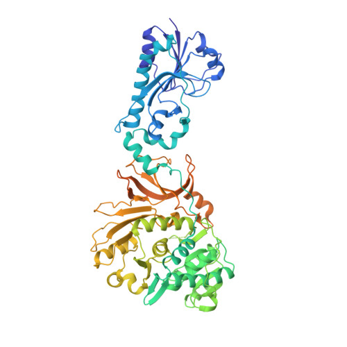
5ES7

Crystal structure of the F-A domains of the LgrA initiation module soaked with FON, AMPcPP, and valine.


Experimental Data Snapshot

  • Method: X-RAY DIFFRACTION
  • Resolution: 2.81 Å
  • R-Value Free: 0.258 
  • R-Value Work: 0.226 
  • R-Value Observed: 0.227 

Starting Model: experimental
View more details

wwPDB Validation   3D Report Full Report


Ligand Structure Quality Assessment 


This is version 1.3 of the entry. See complete history


Literature

Synthetic cycle of the initiation module of a formylating nonribosomal peptide synthetase.

Reimer, J.M.Aloise, M.N.Harrison, P.M.Schmeing, T.M.

(2016) Nature 529: 239-242

  • DOI: https://doi.org/10.1038/nature16503
  • Primary Citation of Related Structures:  
    5ES5, 5ES6, 5ES7, 5ES8, 5ES9

  • PubMed Abstract: 

    Nonribosomal peptide synthetases (NRPSs) are very large proteins that produce small peptide molecules with wide-ranging biological activities, including environmentally friendly chemicals and many widely used therapeutics. NRPSs are macromolecular machines, with modular assembly-line logic, a complex catalytic cycle, moving parts and many active sites. In addition to the core domains required to link the substrates, they often include specialized tailoring domains, which introduce chemical modifications and allow the product to access a large expanse of chemical space. It is still unknown how the NRPS tailoring domains are structurally accommodated into megaenzymes or how they have adapted to function in nonribosomal peptide synthesis. Here we present a series of crystal structures of the initiation module of an antibiotic-producing NRPS, linear gramicidin synthetase. This module includes the specialized tailoring formylation domain, and states are captured that represent every major step of the assembly-line synthesis in the initiation module. The transitions between conformations are large in scale, with both the peptidyl carrier protein domain and the adenylation subdomain undergoing huge movements to transport substrate between distal active sites. The structures highlight the great versatility of NRPSs, as small domains repurpose and recycle their limited interfaces to interact with their various binding partners. Understanding tailoring domains is important if NRPSs are to be utilized in the production of novel therapeutics.


  • Organizational Affiliation

    Department of Biochemistry, McGill University, 3649 Promenade Sir-William-Osler, Montréal, Québec H3G 0B1, Canada.


Macromolecules
Find similar proteins by:  (by identity cutoff)  |  3D Structure
Entity ID: 1
MoleculeChains Sequence LengthOrganismDetailsImage
Linear gramicidin synthetase subunit A685Brevibacillus parabrevisMutation(s): 0 
Gene Names: lgrA
UniProt
Find proteins for Q70LM7 (Brevibacillus parabrevis)
Explore Q70LM7 
Go to UniProtKB:  Q70LM7
Entity Groups  
Sequence Clusters30% Identity50% Identity70% Identity90% Identity95% Identity100% Identity
UniProt GroupQ70LM7
Sequence Annotations
Expand
  • Reference Sequence
Small Molecules
Ligands 3 Unique
IDChains Name / Formula / InChI Key2D Diagram3D Interactions
APC
Query on APC

Download Ideal Coordinates CCD File 
D [auth A]DIPHOSPHOMETHYLPHOSPHONIC ACID ADENOSYL ESTER
C11 H18 N5 O12 P3
CAWZRIXWFRFUQB-IOSLPCCCSA-N
FON
Query on FON

Download Ideal Coordinates CCD File 
B [auth A]N-{[4-({[(6R)-2-amino-5-formyl-4-oxo-1,4,5,6,7,8-hexahydropteridin-6-yl]methyl}amino)phenyl]carbonyl}-L-glutamic acid
C20 H23 N7 O7
VVIAGPKUTFNRDU-OLZOCXBDSA-N
VAL
Query on VAL

Download Ideal Coordinates CCD File 
C [auth A]VALINE
C5 H11 N O2
KZSNJWFQEVHDMF-BYPYZUCNSA-N
Experimental Data & Validation

Experimental Data

  • Method: X-RAY DIFFRACTION
  • Resolution: 2.81 Å
  • R-Value Free: 0.258 
  • R-Value Work: 0.226 
  • R-Value Observed: 0.227 
  • Space Group: P 41 21 2
Unit Cell:
Length ( Å )Angle ( ˚ )
a = 160.943α = 90
b = 160.943β = 90
c = 137.581γ = 90
Software Package:
Software NamePurpose
PHENIXrefinement
HKL-2000data scaling
PDB_EXTRACTdata extraction
HKL-2000data reduction
PHASERphasing

Structure Validation

View Full Validation Report


Ligand Structure Quality Assessment 


Entry History & Funding Information

Deposition Data


Funding OrganizationLocationGrant Number
Canadian Institutes of Health Research (CIHR)Canada106615

Revision History  (Full details and data files)

  • Version 1.0: 2016-01-20
    Type: Initial release
  • Version 1.1: 2016-01-27
    Changes: Database references
  • Version 1.2: 2020-01-08
    Changes: Author supporting evidence, Database references, Derived calculations
  • Version 1.3: 2023-09-27
    Changes: Data collection, Database references, Refinement description