5EQU | pdb_00005equ

Crystal structure of the epimerase SnoN in complex with Fe3+, alpha ketoglutarate and nogalamycin RO


Experimental Data Snapshot

  • Method: X-RAY DIFFRACTION
  • Resolution: 2.20 Å
  • R-Value Free: 
    0.238 (Depositor), 0.245 (DCC) 
  • R-Value Work: 
    0.199 (Depositor), 0.209 (DCC) 
  • R-Value Observed: 
    0.201 (Depositor) 

Starting Model: experimental
View more details

wwPDB Validation 3D Report Full Report

Validation slider image for 5EQU

Ligand Structure Quality Assessment 


This is version 1.2 of the entry. See complete history

Literature

Divergent non-heme iron enzymes in the nogalamycin biosynthetic pathway.

Siitonen, V.Selvaraj, B.Niiranen, L.Lindqvist, Y.Schneider, G.Metsa-Ketela, M.

(2016) Proc Natl Acad Sci U S A 113: 5251-5256

  • DOI: https://doi.org/10.1073/pnas.1525034113
  • Primary Citation Related Structures: 
    5EP9, 5EPA, 5EQU, 5ERL

  • PubMed Abstract: 

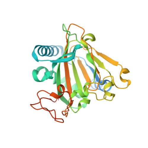
    Nogalamycin, an aromatic polyketide displaying high cytotoxicity, has a unique structure, with one of the carbohydrate units covalently attached to the aglycone via an additional carbon-carbon bond. The underlying chemistry, which implies a particularly challenging reaction requiring activation of an aliphatic carbon atom, has remained enigmatic. Here, we show that the unusual C5''-C2 carbocyclization is catalyzed by the non-heme iron α-ketoglutarate (α-KG)-dependent SnoK in the biosynthesis of the anthracycline nogalamycin. The data are consistent with a mechanistic proposal whereby the Fe(IV) = O center abstracts the H5'' atom from the amino sugar of the substrate, with subsequent attack of the aromatic C2 carbon on the radical center. We further show that, in the same metabolic pathway, the homologous SnoN (38% sequence identity) catalyzes an epimerization step at the adjacent C4'' carbon, most likely via a radical mechanism involving the Fe(IV) = O center. SnoK and SnoN have surprisingly similar active site architectures considering the markedly different chemistries catalyzed by the enzymes. Structural studies reveal that the differences are achieved by minor changes in the alignment of the substrates in front of the reactive ferryl-oxo species. Our findings significantly expand the repertoire of reactions reported for this important protein family and provide an illustrative example of enzyme evolution.


  • Organizational Affiliation
    • Department of Biochemistry, University of Turku, FIN-20014, Turku, Finland;

Macromolecule Content 

  • Total Structure Weight: 135.92 kDa 
  • Atom Count: 8,337 
  • Modeled Residue Count: 1,024 
  • Deposited Residue Count: 1,220 
  • Unique protein chains: 1

Macromolecules

Find similar proteins by:|  3D Structure
Entity ID: 1
MoleculeChains  Sequence LengthOrganismDetailsImage
SnoN,SnoN
A, B, C, D
305Streptomyces nogalaterMutation(s): 0 
Gene Names: snoN
UniProt
Find proteins for Q9EYI0 (Streptomyces nogalater)
Explore Q9EYI0 
Go to UniProtKB:  Q9EYI0
Find proteins for Q9RN67 (Streptomyces nogalater)
Explore Q9RN67 
Go to UniProtKB:  Q9RN67
Entity Groups
Sequence Clusters30% Identity50% Identity70% Identity90% Identity95% Identity100% Identity
UniProt GroupsQ9RN67Q9EYI0
Sequence Annotations
Expand
Reference Sequence

Small Molecules

Ligands 3 Unique
IDChains Name / Formula / InChI Key2D Diagram3D Interactions
5R6

Query on 5R6


Download:Ideal Coordinates CCD File
G [auth A],
J [auth B],
M [auth C],
P [auth D]
Nogalamycin RO
C35 H41 N O14
CRWWSIJFHVGVFW-PDUKGOLSSA-N
AKG

Query on AKG


Download:Ideal Coordinates CCD File
F [auth A],
I [auth B],
L [auth C],
O [auth D]
2-OXOGLUTARIC ACID
C5 H6 O5
KPGXRSRHYNQIFN-UHFFFAOYSA-N
FE

Query on FE


Download:Ideal Coordinates CCD File
E [auth A],
H [auth B],
K [auth C],
N [auth D]
FE (III) ION
Fe
VTLYFUHAOXGGBS-UHFFFAOYSA-N

Experimental Data & Validation

Experimental Data

  • Method: X-RAY DIFFRACTION
  • Resolution: 2.20 Å
  • R-Value Free:  0.238 (Depositor), 0.245 (DCC) 
  • R-Value Work:  0.199 (Depositor), 0.209 (DCC) 
  • R-Value Observed: 0.201 (Depositor) 
Space Group: P 21 21 21
Unit Cell:
Length ( Å )Angle ( ˚ )
a = 71.908α = 90
b = 119.742β = 90
c = 159.447γ = 90
Software Package:
Software NamePurpose
REFMACrefinement
XDSdata reduction
Aimlessdata scaling
PHASERphasing

Structure Validation

View Full Validation Report


Ligand Structure Quality Assessment 


Entry History 

& Funding Information

Deposition Data


Funding OrganizationLocationGrant Number
Swedish Research CouncilSweden--
Finnish AcademyFinland--

Revision History  (Full details and data files)

  • Version 1.0: 2016-05-11
    Type: Initial release
  • Version 1.1: 2016-05-18
    Changes: Database references
  • Version 1.2: 2024-01-10
    Changes: Data collection, Database references, Derived calculations, Refinement description