


Structural dynamics of UDP-galactopyranose mutase from Mycobacterium smegmatis
Wangkanont, K., Kiessling, L.L., Forest, K.T.To be published.
Experimental Data Snapshot
Starting Model: experimental
View more details
Entity ID: 1 | |||||
|---|---|---|---|---|---|
| Molecule | Chains | Sequence Length | Organism | Details | Image |
| UDP-galactopyranose mutase | 400 | Mycolicibacterium smegmatis MC2 155 | Mutation(s): 0 Gene Names: glf, MSMEG_6404, MSMEI_6236 EC: 5.4.99.9 | 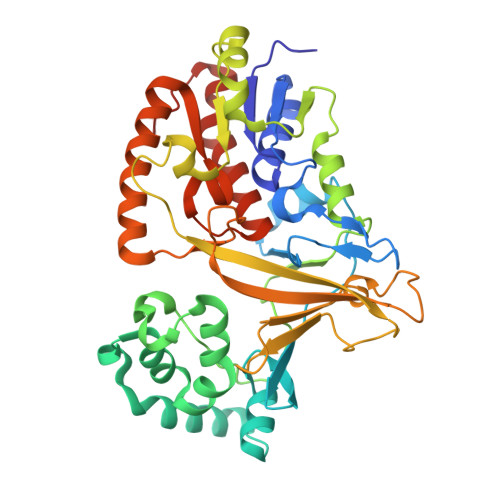 | |
UniProt | |||||
Entity Groups | |||||
| Sequence Clusters | 30% Identity50% Identity70% Identity90% Identity95% Identity100% Identity | ||||
| UniProt Group | A0R629 | ||||
Sequence AnnotationsExpand | |||||
Reference Sequence | |||||
| Ligands 4 Unique | |||||
|---|---|---|---|---|---|
| ID | Chains | Name / Formula / InChI Key | 2D Diagram | 3D Interactions | |
| FAD Download:Ideal Coordinates CCD File | C [auth A], K [auth B] | FLAVIN-ADENINE DINUCLEOTIDE C27 H33 N9 O15 P2 VWWQXMAJTJZDQX-UYBVJOGSSA-N |  | ||
| UDP Download:Ideal Coordinates CCD File | J [auth B] | URIDINE-5'-DIPHOSPHATE C9 H14 N2 O12 P2 XCCTYIAWTASOJW-XVFCMESISA-N |  | ||
| SO4 Download:Ideal Coordinates CCD File | F [auth A] H [auth A] O [auth B] P [auth B] Q [auth B] | SULFATE ION O4 S QAOWNCQODCNURD-UHFFFAOYSA-L |  | ||
| NO3 Download:Ideal Coordinates CCD File | D [auth A] E [auth A] G [auth A] I [auth A] L [auth B] | NITRATE ION N O3 NHNBFGGVMKEFGY-UHFFFAOYSA-N |  | ||
| Length ( Å ) | Angle ( ˚ ) |
|---|---|
| a = 124.188 | α = 90 |
| b = 132.184 | β = 90 |
| c = 135.654 | γ = 90 |
| Software Name | Purpose |
|---|---|
| SCALEPACK | data scaling |
| PHASER | phasing |
| PHENIX | refinement |
| PDB_EXTRACT | data extraction |
| HKL-2000 | data reduction |
| Funding Organization | Location | Grant Number |
|---|---|---|
| National Institutes of Health/National Institute Of Allergy and Infectious Diseases (NIH/NIAID) | United States | R01 AI063596 |
| National Institutes of Health/National Institute of General Medical Sciences (NIH/NIGMS) | United States | R01 GM100346 |


