5EP4 | pdb_00005ep4

Structure, Regulation, and Inhibition of the Quorum-Sensing Signal Integrator LuxO


Experimental Data Snapshot

  • Method: X-RAY DIFFRACTION
  • Resolution: 1.50 Å
  • R-Value Free: 
    0.182 (Depositor), 0.184 (DCC) 
  • R-Value Work: 
    0.155 (Depositor), 0.155 (DCC) 
  • R-Value Observed: 
    0.156 (Depositor) 

wwPDB Validation 3D Report Full Report

Validation slider image for 5EP4

Ligand Structure Quality Assessment 


This is version 1.4 of the entry. See complete history

Literature

Structure, Regulation, and Inhibition of the Quorum-Sensing Signal Integrator LuxO.

Boyaci, H.Shah, T.Hurley, A.Kokona, B.Li, Z.Ventocilla, C.Jeffrey, P.D.Semmelhack, M.F.Fairman, R.Bassler, B.L.Hughson, F.M.

(2016) PLoS Biol 14: e1002464-e1002464

  • DOI: https://doi.org/10.1371/journal.pbio.1002464
  • Primary Citation Related Structures: 
    5EP0, 5EP1, 5EP2, 5EP3, 5EP4

  • PubMed Abstract: 

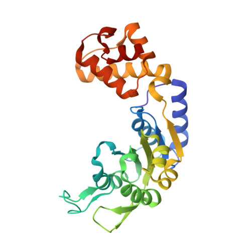
    In a process called quorum sensing, bacteria communicate with chemical signal molecules called autoinducers to control collective behaviors. In pathogenic vibrios, including Vibrio cholerae, the accumulation of autoinducers triggers repression of genes responsible for virulence factor production and biofilm formation. The vibrio autoinducer molecules bind to transmembrane receptors of the two-component histidine sensor kinase family. Autoinducer binding inactivates the receptors' kinase activities, leading to dephosphorylation and inhibition of the downstream response regulator LuxO. Here, we report the X-ray structure of LuxO in its unphosphorylated, autoinhibited state. Our structure reveals that LuxO, a bacterial enhancer-binding protein of the AAA+ ATPase superfamily, is inhibited by an unprecedented mechanism in which a linker that connects the catalytic and regulatory receiver domains occupies the ATPase active site. The conformational change that accompanies receiver domain phosphorylation likely disrupts this interaction, providing a mechanistic rationale for LuxO activation. We also determined the crystal structure of the LuxO catalytic domain bound to a broad-spectrum inhibitor. The inhibitor binds in the ATPase active site and recapitulates elements of the natural regulatory mechanism. Remarkably, a single inhibitor molecule may be capable of inhibiting an entire LuxO oligomer.


  • Organizational Affiliation
    • Department of Molecular Biology, Princeton University, Princeton, New Jersey, United States of America.

Macromolecule Content 

  • Total Structure Weight: 29.42 kDa 
  • Atom Count: 2,311 
  • Modeled Residue Count: 253 
  • Deposited Residue Count: 256 
  • Unique protein chains: 1

Macromolecules

Find similar proteins by:|  3D Structure
Entity ID: 1
MoleculeChains  Sequence LengthOrganismDetailsImage
Putative repressor protein luxO256Photobacterium angustumMutation(s): 0 
UniProt
Find proteins for Q1ZS18 (Photobacterium angustum (strain S14 / CCUG 15956))
Explore Q1ZS18 
Go to UniProtKB:  Q1ZS18
Entity Groups
Sequence Clusters30% Identity50% Identity70% Identity90% Identity95% Identity100% Identity
UniProt GroupQ1ZS18
Sequence Annotations
Expand
Reference Sequence

Small Molecules

Experimental Data & Validation

Experimental Data

  • Method: X-RAY DIFFRACTION
  • Resolution: 1.50 Å
  • R-Value Free:  0.182 (Depositor), 0.184 (DCC) 
  • R-Value Work:  0.155 (Depositor), 0.155 (DCC) 
  • R-Value Observed: 0.156 (Depositor) 
Space Group: P 61
Unit Cell:
Length ( Å )Angle ( ˚ )
a = 73.69α = 90
b = 73.69β = 90
c = 81.792γ = 120
Software Package:
Software NamePurpose
SCALEPACKdata scaling
PHENIXrefinement
PDB_EXTRACTdata extraction
DENZOdata reduction

Structure Validation

View Full Validation Report


Ligand Structure Quality Assessment 


Entry History 

& Funding Information

Deposition Data


Funding OrganizationLocationGrant Number
National Institutes of Health/National Institute Of Allergy and Infectious Diseases (NIH/NIAID)United StatesAI054442

Revision History  (Full details and data files)

  • Version 1.0: 2016-04-20
    Type: Initial release
  • Version 1.1: 2016-06-08
    Changes: Database references
  • Version 1.2: 2017-09-27
    Changes: Author supporting evidence, Derived calculations
  • Version 1.3: 2019-12-11
    Changes: Author supporting evidence
  • Version 1.4: 2024-03-06
    Changes: Data collection, Database references