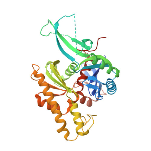
5EOU

Pseudomonas aeruginosa PilM:PilN1-12 bound to ATP


Experimental Data Snapshot

  • Method: X-RAY DIFFRACTION
  • Resolution: 2.40 Å
  • R-Value Free: 0.239 
  • R-Value Work: 0.212 
  • R-Value Observed: 0.214 

Starting Model: experimental
View more details

wwPDB Validation   3D Report Full Report


Ligand Structure Quality Assessment 


This is version 2.0 of the entry. See complete history


Literature

PilN Binding Modulates the Structure and Binding Partners of the Pseudomonas aeruginosa Type IVa Pilus Protein PilM.

McCallum, M.Tammam, S.Little, D.J.Robinson, H.Koo, J.Shah, M.Calmettes, C.Moraes, T.F.Burrows, L.L.Howell, P.L.

(2016) J Biol Chem 291: 11003-11015

  • DOI: https://doi.org/10.1074/jbc.M116.718353
  • Primary Citation of Related Structures:  
    5EOU, 5EOX, 5EOY, 5EQ6

  • PubMed Abstract: 

    Pseudomonas aeruginosa is an opportunistic bacterial pathogen that expresses type IVa pili. The pilus assembly system, which promotes surface-associated twitching motility and virulence, is composed of inner and outer membrane subcomplexes, connected by an alignment subcomplex composed of PilMNOP. PilM binds to the N terminus of PilN, and we hypothesize that this interaction causes functionally significant structural changes in PilM. To characterize this interaction, we determined the crystal structures of PilM and a PilM chimera where PilM was fused to the first 12 residues of PilN (PilM·PilN(1-12)). Structural analysis, multiangle light scattering coupled with size exclusion chromatography, and bacterial two-hybrid data revealed that PilM forms dimers mediated by the binding of a novel conserved motif in the N terminus of PilM, and binding PilN abrogates this binding interface, resulting in PilM monomerization. Structural comparison of PilM with PilM·PilN(1-12) revealed that upon PilN binding, there is a large domain closure in PilM that alters its ATP binding site. Using biolayer interferometry, we found that the association rate of PilN with PilM is higher in the presence of ATP compared with ADP. Bacterial two-hybrid data suggested the connectivity of the cytoplasmic and inner membrane components of the type IVa pilus machinery in P. aeruginosa, with PilM binding to PilB, PilT, and PilC in addition to PilN. Pull-down experiments demonstrated direct interactions of PilM with PilB and PilT. We propose a working model in which dynamic binding of PilN facilitates functionally relevant structural changes in PilM.


  • Organizational Affiliation

    From the Department of Biochemistry, University of Toronto, Toronto, Ontario M5S 1A8, Canada, the Program in Molecular Structure and Function, Peter Gilgan Centre for Research and Learning, Hospital for Sick Children, Toronto, Ontario M5G 0A4, Canada.


Macromolecules
Find similar proteins by:  (by identity cutoff)  |  3D Structure
Entity ID: 1
MoleculeChains Sequence LengthOrganismDetailsImage
Type 4 fimbrial biogenesis protein PilM,Type 4 fimbrial biogenesis protein PilN
A, B
372Pseudomonas aeruginosa PAO1Mutation(s): 0 
Gene Names: pilMPA5044pilNPA5043
UniProt
Find proteins for G3XD28 (Pseudomonas aeruginosa (strain ATCC 15692 / DSM 22644 / CIP 104116 / JCM 14847 / LMG 12228 / 1C / PRS 101 / PAO1))
Explore G3XD28 
Go to UniProtKB:  G3XD28
Find proteins for G3XD30 (Pseudomonas aeruginosa (strain ATCC 15692 / DSM 22644 / CIP 104116 / JCM 14847 / LMG 12228 / 1C / PRS 101 / PAO1))
Explore G3XD30 
Go to UniProtKB:  G3XD30
Entity Groups  
Sequence Clusters30% Identity50% Identity70% Identity90% Identity95% Identity100% Identity
UniProt GroupsG3XD30G3XD28
Sequence Annotations
Expand
  • Reference Sequence
Small Molecules
Ligands 5 Unique
IDChains Name / Formula / InChI Key2D Diagram3D Interactions
ATP
Query on ATP

Download Ideal Coordinates CCD File 
K [auth B]ADENOSINE-5'-TRIPHOSPHATE
C10 H16 N5 O13 P3
ZKHQWZAMYRWXGA-KQYNXXCUSA-N
SO4
Query on SO4

Download Ideal Coordinates CCD File 
C [auth A]
D [auth A]
E [auth A]
F [auth A]
G [auth A]
C [auth A],
D [auth A],
E [auth A],
F [auth A],
G [auth A],
L [auth B],
M [auth B],
N [auth B]
SULFATE ION
O4 S
QAOWNCQODCNURD-UHFFFAOYSA-L
CL
Query on CL

Download Ideal Coordinates CCD File 
H [auth A],
O [auth B],
P [auth B],
Q [auth B]
CHLORIDE ION
Cl
VEXZGXHMUGYJMC-UHFFFAOYSA-M
MG
Query on MG

Download Ideal Coordinates CCD File 
J [auth A],
S [auth B]
MAGNESIUM ION
Mg
JLVVSXFLKOJNIY-UHFFFAOYSA-N
NA
Query on NA

Download Ideal Coordinates CCD File 
I [auth A],
R [auth B]
SODIUM ION
Na
FKNQFGJONOIPTF-UHFFFAOYSA-N
Experimental Data & Validation

Experimental Data

  • Method: X-RAY DIFFRACTION
  • Resolution: 2.40 Å
  • R-Value Free: 0.239 
  • R-Value Work: 0.212 
  • R-Value Observed: 0.214 
  • Space Group: P 3 2 1
Unit Cell:
Length ( Å )Angle ( ˚ )
a = 131.79α = 90
b = 131.79β = 90
c = 78.993γ = 120
Software Package:
Software NamePurpose
PHENIXrefinement
Cootmodel building
HKL-2000data scaling
PHASERphasing
HKL-2000data reduction

Structure Validation

View Full Validation Report


Ligand Structure Quality Assessment 


Entry History 

Deposition Data

Revision History  (Full details and data files)

  • Version 1.0: 2016-04-27
    Type: Initial release
  • Version 1.1: 2016-06-08
    Changes: Database references
  • Version 2.0: 2023-09-27
    Changes: Atomic model, Data collection, Database references, Derived calculations, Refinement description