5EOM | pdb_00005eom

Structure of full-length human MAB21L1 with bound CTP


Experimental Data Snapshot

  • Method: X-RAY DIFFRACTION
  • Resolution: 2.55 Å
  • R-Value Free: 
    0.227 (Depositor), 0.234 (DCC) 
  • R-Value Work: 
    0.199 (Depositor), 0.207 (DCC) 
  • R-Value Observed: 
    0.200 (Depositor) 

Starting Model: experimental
View more details

wwPDB Validation 3D Report Full Report

Validation slider image for 5EOM

Ligand Structure Quality Assessment 


This is version 1.2 of the entry. See complete history

Literature

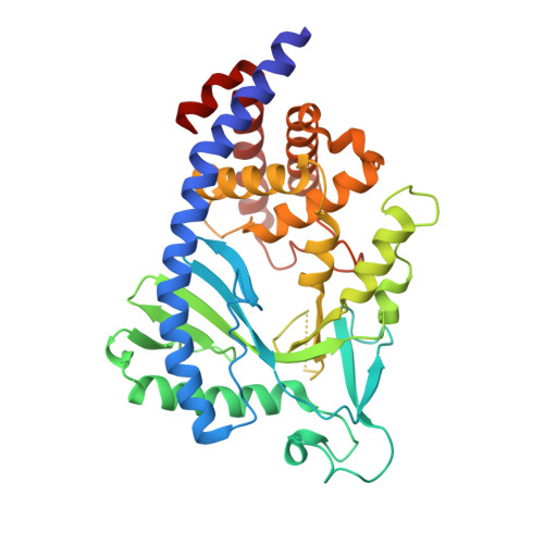
Structural and biochemical characterization of the cell fate determining nucleotidyltransferase fold protein MAB21L1.

de Oliveira Mann, C.C.Kiefersauer, R.Witte, G.Hopfner, K.P.

(2016) Sci Rep 6: 27498-27498

  • DOI: https://doi.org/10.1038/srep27498
  • Primary Citation Related Structures: 
    5EOG, 5EOM

  • PubMed Abstract: 

    The exceptionally conserved metazoan MAB21 proteins are implicated in cell fate decisions and share considerable sequence homology with the cyclic GMP-AMP synthase. cGAS is the major innate immune sensor for cytosolic DNA and produces the second messenger 2'-5', 3'-5' cyclic GMP-AMP. Little is known about the structure and biochemical function of other proteins of the cGAS-MAB21 subfamily, such as MAB21L1, MAB21L2 and MAB21L3. We have determined the crystal structure of human full-length MAB21L1. Our analysis reveals high structural conservation between MAB21L1 and cGAS but also uncovers important differences. Although monomeric in solution, MAB21L1 forms a highly symmetric double-pentameric oligomer in the crystal, raising the possibility that oligomerization could be a feature of MAB21L1. In the crystal, MAB21L1 is in an inactive conformation requiring a conformational change - similar to cGAS - to develop any nucleotidyltransferase activity. Co-crystallization with NTP identified a putative ligand binding site of MAB21 proteins that corresponds to the DNA binding site of cGAS. Finally, we offer a structure-based explanation for the effects of MAB21L2 mutations in patients with eye malformations. The underlying residues participate in fold-stabilizing interaction networks and mutations destabilize the protein. In summary, we provide a first structural framework for MAB21 proteins.


  • Organizational Affiliation
    • Ludwig-Maximilians-Universität München, Gene Center and Dept. of Biochemistry, Feodor-Lynen-Str. 25, 81377 Munich, Germany.

Macromolecule Content 

  • Total Structure Weight: 419.65 kDa 
  • Atom Count: 28,115 
  • Modeled Residue Count: 3,409 
  • Deposited Residue Count: 3,620 
  • Unique protein chains: 1

Macromolecules

Find similar proteins by:|  3D Structure
Entity ID: 1
MoleculeChains  Sequence LengthOrganismDetailsImage
Protein mab-21-like 1362Homo sapiensMutation(s): 0 
Gene Names: MAB21L1CAGR1Nbla00126
EC: 2.7.7
UniProt & NIH Common Fund Data Resources
Find proteins for Q13394 (Homo sapiens)
Explore Q13394 
Go to UniProtKB:  Q13394
PHAROS:  Q13394
GTEx:  ENSG00000180660 
Entity Groups
Sequence Clusters30% Identity50% Identity70% Identity90% Identity95% Identity100% Identity
UniProt GroupQ13394
Sequence Annotations
Expand
Reference Sequence

Small Molecules

Ligands 3 Unique
IDChains Name / Formula / InChI Key2D Diagram3D Interactions
CTP

Query on CTP


Download:Ideal Coordinates CCD File
AA [auth H]
CA [auth I]
DA [auth J]
HA [auth C]
M [auth A]
AA [auth H],
CA [auth I],
DA [auth J],
HA [auth C],
M [auth A],
O [auth B],
Q [auth D],
S [auth E],
V [auth F],
X [auth G]
CYTIDINE-5'-TRIPHOSPHATE
C9 H16 N3 O14 P3
PCDQPRRSZKQHHS-XVFCMESISA-N
CIT

Query on CIT


Download:Ideal Coordinates CCD File
BA [auth I]
EA [auth C]
K [auth A]
N [auth B]
P [auth D]
BA [auth I],
EA [auth C],
K [auth A],
N [auth B],
P [auth D],
R [auth E],
T [auth F],
W [auth G],
Y [auth H],
Z [auth H]
CITRIC ACID
C6 H8 O7
KRKNYBCHXYNGOX-UHFFFAOYSA-N
TMO

Query on TMO


Download:Ideal Coordinates CCD File
FA [auth C],
GA [auth C],
L [auth A],
U [auth F]
trimethylamine oxide
C3 H9 N O
UYPYRKYUKCHHIB-UHFFFAOYSA-N

Experimental Data & Validation

Experimental Data

  • Method: X-RAY DIFFRACTION
  • Resolution: 2.55 Å
  • R-Value Free:  0.227 (Depositor), 0.234 (DCC) 
  • R-Value Work:  0.199 (Depositor), 0.207 (DCC) 
  • R-Value Observed: 0.200 (Depositor) 
Space Group: P 1 21 1
Unit Cell:
Length ( Å )Angle ( ˚ )
a = 115α = 90
b = 177.76β = 97.58
c = 134.94γ = 90
Software Package:
Software NamePurpose
PHENIXrefinement
XDSdata reduction
XSCALEdata scaling
PHASERphasing

Structure Validation

View Full Validation Report


Ligand Structure Quality Assessment 


Entry History 

& Funding Information

Deposition Data


Funding OrganizationLocationGrant Number
German Research FoundationGermanyGRK1721

Revision History  (Full details and data files)

  • Version 1.0: 2016-06-01
    Type: Initial release
  • Version 1.1: 2016-06-22
    Changes: Database references
  • Version 1.2: 2024-05-01
    Changes: Author supporting evidence, Data collection, Database references, Refinement description