5EMJ | pdb_00005emj

Crystal structure of PRMT5:MEP50 with Compound 8 and sinefungin


Experimental Data Snapshot

  • Method: X-RAY DIFFRACTION
  • Resolution: 2.27 Å
  • R-Value Free: 
    0.266 (Depositor), 0.265 (DCC) 
  • R-Value Work: 
    0.209 (Depositor), 0.208 (DCC) 
  • R-Value Observed: 
    0.211 (Depositor) 

wwPDB Validation 3D Report Full Report

Validation slider image for 5EMJ

Ligand Structure Quality Assessment 


This is version 1.3 of the entry. See complete history

Literature

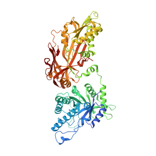
Structure and Property Guided Design in the Identification of PRMT5 Tool Compound EPZ015666.

Duncan, K.W.Rioux, N.Boriack-Sjodin, P.A.Munchhof, M.J.Reiter, L.A.Majer, C.R.Jin, L.Johnston, L.D.Chan-Penebre, E.Kuplast, K.G.Porter Scott, M.Pollock, R.M.Waters, N.J.Smith, J.J.Moyer, M.P.Copeland, R.A.Chesworth, R.

(2016) ACS Med Chem Lett 7: 162-166

  • DOI: https://doi.org/10.1021/acsmedchemlett.5b00380
  • Primary Citation Related Structures: 
    5EMJ, 5EMK, 5EML, 5EMM

  • PubMed Abstract: 

    The recent publication of a potent and selective inhibitor of protein methyltransferase 5 (PRMT5) provides the scientific community with in vivo-active tool compound EPZ015666 (GSK3235025) to probe the underlying pharmacology of this key enzyme. Herein, we report the design and optimization strategies employed on an initial hit compound with poor in vitro clearance to yield in vivo tool compound EPZ015666 and an additional potent in vitro tool molecule EPZ015866 (GSK3203591).


  • Organizational Affiliation
    • Epizyme, Inc. , 400 Technology Square, Cambridge, Massachusetts 02139, United States.

Macromolecule Content 

  • Total Structure Weight: 112.79 kDa 
  • Atom Count: 7,774 
  • Modeled Residue Count: 928 
  • Deposited Residue Count: 995 
  • Unique protein chains: 2

Macromolecules

Find similar proteins by:|  3D Structure
Entity ID: 1
MoleculeChains  Sequence LengthOrganismDetailsImage
Protein arginine N-methyltransferase 5645Homo sapiensMutation(s): 0 
Gene Names: PRMT5HRMT1L5IBP72JBP1SKB1
EC: 2.1.1 (PDB Primary Data), 2.1.1.125 (PDB Primary Data), 2.1.1.320 (UniProt)
UniProt & NIH Common Fund Data Resources
Find proteins for O14744 (Homo sapiens)
Explore O14744 
Go to UniProtKB:  O14744
PHAROS:  O14744
GTEx:  ENSG00000100462 
Entity Groups
Sequence Clusters30% Identity50% Identity70% Identity90% Identity95% Identity100% Identity
UniProt GroupO14744
Sequence Annotations
Expand
Reference Sequence
Find similar proteins by:|  3D Structure
Entity ID: 2
MoleculeChains  Sequence LengthOrganismDetailsImage
Methylosome protein 50350Homo sapiensMutation(s): 0 
Gene Names: WDR77MEP50WD45HKMT1069Nbla10071
UniProt & NIH Common Fund Data Resources
Find proteins for Q9BQA1 (Homo sapiens)
Explore Q9BQA1 
Go to UniProtKB:  Q9BQA1
PHAROS:  Q9BQA1
GTEx:  ENSG00000116455 
Entity Groups
Sequence Clusters30% Identity50% Identity70% Identity90% Identity95% Identity100% Identity
UniProt GroupQ9BQA1
Sequence Annotations
Expand
Reference Sequence

Small Molecules

Ligands 3 Unique
IDChains Name / Formula / InChI Key2D Diagram3D Interactions
5QJ

Query on 5QJ


Download:Ideal Coordinates CCD File
D [auth A](2~{S})-1-(3,4-dihydro-1~{H}-isoquinolin-2-yl)-3-[[4-(3-methylbenzimidazol-5-yl)pyridin-2-yl]amino]propan-2-ol
C25 H27 N5 O
PNOIRPBZBNYURC-QFIPXVFZSA-N
SFG

Query on SFG


Download:Ideal Coordinates CCD File
C [auth A]SINEFUNGIN
C15 H23 N7 O5
LMXOHSDXUQEUSF-YECHIGJVSA-N
GOL

Query on GOL


Download:Ideal Coordinates CCD File
E [auth A],
F [auth A],
G [auth A],
H [auth B]
GLYCEROL
C3 H8 O3
PEDCQBHIVMGVHV-UHFFFAOYSA-N

Experimental Data & Validation

Experimental Data

  • Method: X-RAY DIFFRACTION
  • Resolution: 2.27 Å
  • R-Value Free:  0.266 (Depositor), 0.265 (DCC) 
  • R-Value Work:  0.209 (Depositor), 0.208 (DCC) 
  • R-Value Observed: 0.211 (Depositor) 
Space Group: I 2 2 2
Unit Cell:
Length ( Å )Angle ( ˚ )
a = 104.24α = 90
b = 138.83β = 90
c = 179.34γ = 90
Software Package:
Software NamePurpose
SCALAdata scaling
REFMACrefinement
PDB_EXTRACTdata extraction
iMOSFLMdata reduction

Structure Validation

View Full Validation Report


Ligand Structure Quality Assessment 


Entry History 

Deposition Data

Revision History  (Full details and data files)

  • Version 1.0: 2016-02-24
    Type: Initial release
  • Version 1.1: 2016-07-27
    Changes: Data collection
  • Version 1.2: 2018-04-18
    Changes: Data collection, Database references, Derived calculations
  • Version 1.3: 2024-10-23
    Changes: Data collection, Database references, Structure summary