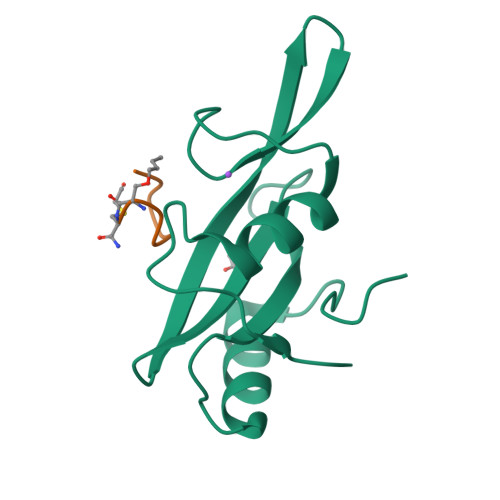
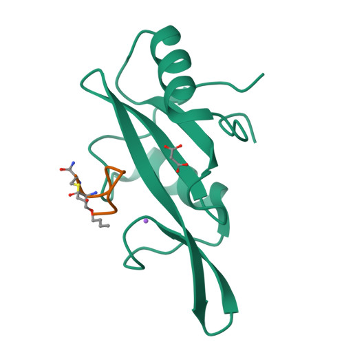
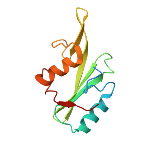
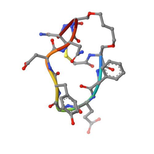
5EEL

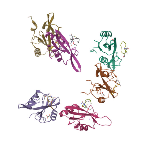
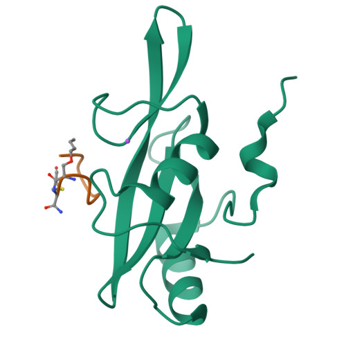
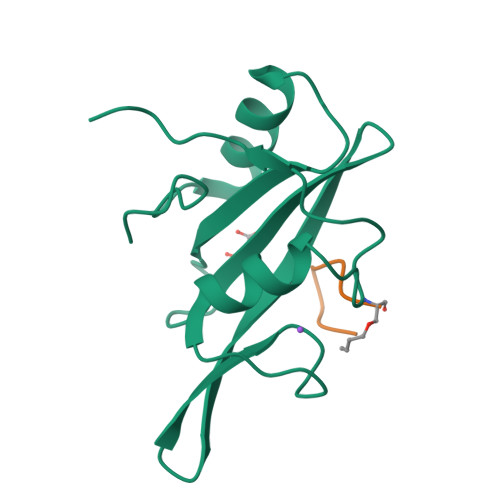
Grb7 SH2 with bicyclic peptide inhibitor


Experimental Data Snapshot

  • Method: X-RAY DIFFRACTION
  • Resolution: 2.47 Å
  • R-Value Free: 0.232 
  • R-Value Work: 0.185 
  • R-Value Observed: 0.188 

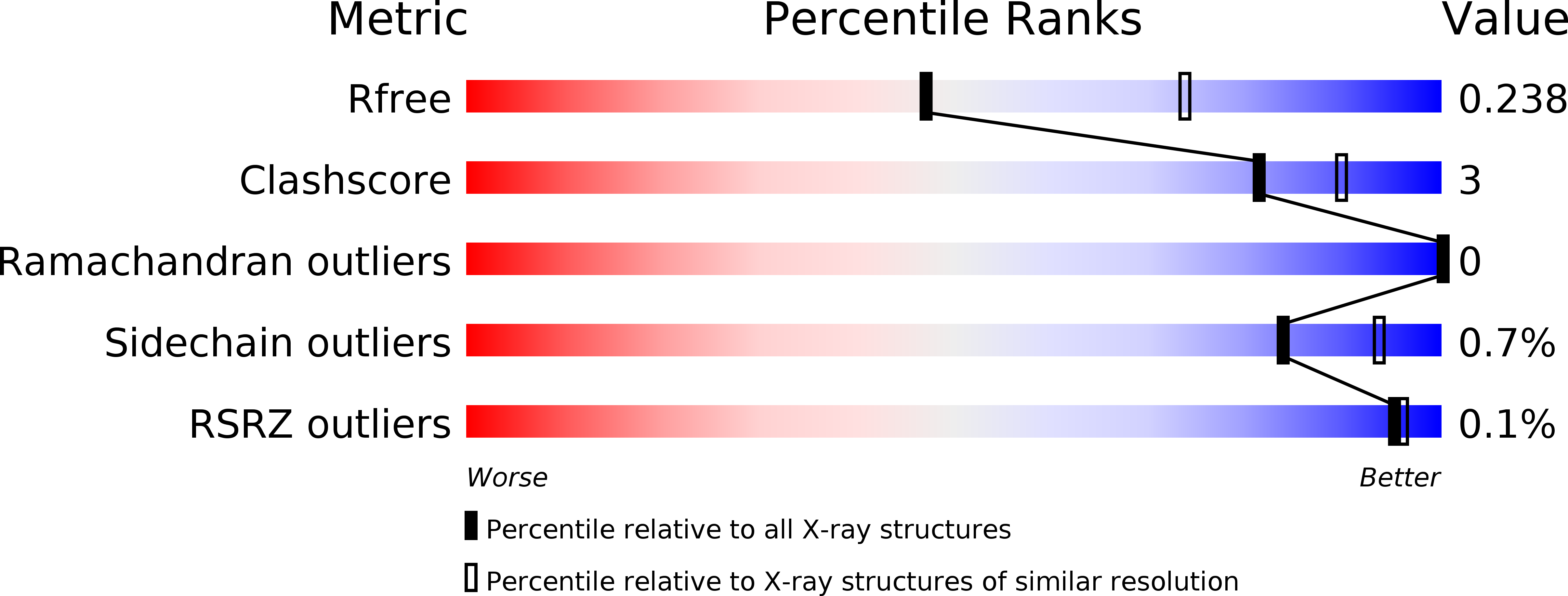
wwPDB Validation   3D Report Full Report


This is version 2.0 of the entry. See complete history


Literature

Unexpected involvement of staple leads to redesign of selective bicyclic peptide inhibitor of Grb7.

Gunzburg, M.J.Kulkarni, K.Watson, G.M.Ambaye, N.D.Del Borgo, M.P.Brandt, R.Pero, S.C.Perlmutter, P.Wilce, M.C.Wilce, J.A.

(2016) Sci Rep 6: 27060-27060

  • DOI: https://doi.org/10.1038/srep27060
  • Primary Citation of Related Structures:  
    5D0J, 5EEL, 5EEQ

  • PubMed Abstract: 

    The design of potent and specific peptide inhibitors to therapeutic targets is of enormous utility for both proof-of-concept studies and for the development of potential new therapeutics. Grb7 is a key signaling molecule in the progression of HER2 positive and triple negative breast cancers. Here we report the crystal structure of a stapled bicyclic peptide inhibitor G7-B1 in complex with the Grb7-SH2 domain. This revealed an unexpected binding mode of the peptide, in which the staple forms an alternative contact with the surface of the target protein. Based on this structural information, we designed a new series of bicyclic G7 peptides that progressively constrain the starting peptide, to arrive at the G7-B4 peptide that binds with an approximately 2-fold enhanced affinity to the Grb7-SH2 domain (KD = 0.83 μM) compared to G7-B1 and shows low affinity binding to Grb2-, Grb10- and Grb14-SH2 domains (KD > 100 μM). Furthermore, we determined the structure of the G7-B4 bicyclic peptide in complex with the Grb7-SH2 domain, both before and after ring closing metathesis to show that the closed staple is essential to the target interaction. The G7-B4 peptide represents an advance in the development of Grb7 inhibitors and is a classical example of structure aided inhibitor development.


  • Organizational Affiliation

    Biomedicine Discovery Institute, Department of Biochemistry and Molecular Biology, Monash University, Wellington Road, Clayton VIC 3800, Australia.


Macromolecules
Find similar proteins by:  (by identity cutoff)  |  3D Structure
Entity ID: 1
MoleculeChains Sequence LengthOrganismDetailsImage
Growth factor receptor-bound protein 7
A, B, C, D, E
A, B, C, D, E, F
120Homo sapiensMutation(s): 0 
Gene Names: GRB7
UniProt & NIH Common Fund Data Resources
Find proteins for Q14451 (Homo sapiens)
Explore Q14451 
Go to UniProtKB:  Q14451
PHAROS:  Q14451
GTEx:  ENSG00000141738 
Entity Groups  
Sequence Clusters30% Identity50% Identity70% Identity90% Identity95% Identity100% Identity
UniProt GroupQ14451
Sequence Annotations
Expand
  • Reference Sequence

Find similar proteins by:  Sequence   |   3D Structure  

Entity ID: 2
MoleculeChains Sequence LengthOrganismDetailsImage
Bicyclic Peptide Inhibitor9synthetic constructMutation(s): 0 
Sequence Annotations
Expand
  • Reference Sequence
Small Molecules
Ligands 3 Unique
IDChains Name / Formula / InChI Key2D Diagram3D Interactions
MLA
Query on MLA

Download Ideal Coordinates CCD File 
AA [auth F],
M [auth A],
R [auth C],
U [auth D],
X [auth E]
MALONIC ACID
C3 H4 O4
OFOBLEOULBTSOW-UHFFFAOYSA-N
FMT
Query on FMT

Download Ideal Coordinates CCD File 
BA [auth F]
N [auth A]
P [auth B]
S [auth C]
V [auth D]
BA [auth F],
N [auth A],
P [auth B],
S [auth C],
V [auth D],
Y [auth E]
FORMIC ACID
C H2 O2
BDAGIHXWWSANSR-UHFFFAOYSA-N
NA
Query on NA

Download Ideal Coordinates CCD File 
CA [auth F]
O [auth A]
Q [auth B]
T [auth C]
W [auth D]
CA [auth F],
O [auth A],
Q [auth B],
T [auth C],
W [auth D],
Z [auth E]
SODIUM ION
Na
FKNQFGJONOIPTF-UHFFFAOYSA-N
Modified Residues  2 Unique
IDChains TypeFormula2D DiagramParent
73C
Query on 73C
G [auth L]
H [auth M]
I [auth N]
J [auth P]
K [auth Q]
G [auth L],
H [auth M],
I [auth N],
J [auth P],
K [auth Q],
L [auth R]
L-PEPTIDE LINKINGC7 H15 N O3SER
AEA
Query on AEA
G [auth L]
H [auth M]
I [auth N]
J [auth P]
K [auth Q]
G [auth L],
H [auth M],
I [auth N],
J [auth P],
K [auth Q],
L [auth R]
L-PEPTIDE LINKINGC5 H10 N2 O3 SCYS
Experimental Data & Validation

Experimental Data

  • Method: X-RAY DIFFRACTION
  • Resolution: 2.47 Å
  • R-Value Free: 0.232 
  • R-Value Work: 0.185 
  • R-Value Observed: 0.188 
  • Space Group: P 41 21 2
Unit Cell:
Length ( Å )Angle ( ˚ )
a = 95.228α = 90
b = 95.228β = 90
c = 241.566γ = 90
Software Package:
Software NamePurpose
PHENIXrefinement
PDB_EXTRACTdata extraction
xia2data reduction
xia2data scaling
MOLREPphasing

Structure Validation

View Full Validation Report


Entry History 

Deposition Data

Revision History  (Full details and data files)

  • Version 1.0: 2016-06-15
    Type: Initial release
  • Version 2.0: 2018-12-26
    Changes: Data collection, Derived calculations, Polymer sequence