5E9L | pdb_00005e9l

Crystal Structure of BAZ2B bromodomain in complex with fragment F103


Experimental Data Snapshot

  • Method: X-RAY DIFFRACTION
  • Resolution: 1.90 Å
  • R-Value Free: 
    0.212 (Depositor), 0.222 (DCC) 
  • R-Value Work: 
    0.179 (Depositor), 0.189 (DCC) 
  • R-Value Observed: 
    0.181 (Depositor) 

Starting Model: experimental
View more details

wwPDB Validation 3D Report Full Report

Validation slider image for 5E9L

This is version 1.2 of the entry. See complete history

Literature

High-Throughput Fragment Docking into the BAZ2B Bromodomain: Efficient in Silico Screening for X-Ray Crystallography.

Lolli, G.Caflisch, A.

(2016) ACS Chem Biol 11: 800-807

  • DOI: https://doi.org/10.1021/acschembio.5b00914
  • Primary Citation Related Structures: 
    5DYU, 5DYX, 5E9I, 5E9K, 5E9L, 5E9M, 5E9Y, 5EA1

  • PubMed Abstract: 

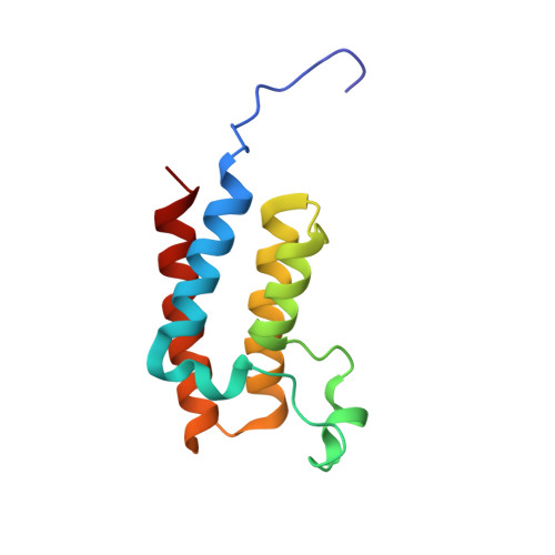
    Bromodomains are protein modules that bind to acetylated lysine side chains in histones and other proteins. The bromodomain adjacent to zinc finger domain protein 2B (BAZ2B) has been reported to be poorly druggable. Here, we screened an in-house library of 350 fragments by automatic docking to the BAZ2B bromodomain. The top 12 fragments according to the predicted binding energy were selected for experiments of soaking into apo crystals of BAZ2B which yielded the structure of the complex for four of them, which is a hit rate of 33%. Additional crystal structures were solved for BAZ2B and two scaffolds identified by analogy. For three topologically similar fragments, the crystal structures reveal binding modes with different penetration, i.e., with zero, one, and two water molecules, respectively, located between the fragment and the side chain of a conserved tyrosine (Tyr1901) in the bottom of the acetyl lysine pocket of BAZ2B. Furthermore, a remarkable stereoselectivity of the acetyl lysine pocket emerges from the crystal structures of the bromodomains of BAZ2B and SMARCA4 in complex with the chiral diol MPD (2-methyl-2,4-pentanediol).


  • Organizational Affiliation
    • Department of Biochemistry, University of Zürich , Winterthurerstrasse 190, CH-8057, Zürich, Switzerland.

Macromolecule Content 

  • Total Structure Weight: 13.66 kDa 
  • Atom Count: 1,095 
  • Modeled Residue Count: 116 
  • Deposited Residue Count: 116 
  • Unique protein chains: 1

Macromolecules

Find similar proteins by:|  3D Structure
Entity ID: 1
MoleculeChains  Sequence LengthOrganismDetailsImage
Bromodomain adjacent to zinc finger domain protein 2B116Homo sapiensMutation(s): 0 
Gene Names: BAZ2BKIAA1476
UniProt & NIH Common Fund Data Resources
Find proteins for Q9UIF8 (Homo sapiens)
Explore Q9UIF8 
Go to UniProtKB:  Q9UIF8
PHAROS:  Q9UIF8
GTEx:  ENSG00000123636 
Entity Groups
Sequence Clusters30% Identity50% Identity70% Identity90% Identity95% Identity100% Identity
UniProt GroupQ9UIF8
Sequence Annotations
Expand
Reference Sequence

Small Molecules

Ligands 1 Unique
IDChains Name / Formula / InChI Key2D Diagram3D Interactions
6AI

Query on 6AI


Download:Ideal Coordinates CCD File
B [auth A]1H-indazol-6-amine
C7 H7 N3
KEJFADGISRFLFO-UHFFFAOYSA-N

Experimental Data & Validation

Experimental Data

  • Method: X-RAY DIFFRACTION
  • Resolution: 1.90 Å
  • R-Value Free:  0.212 (Depositor), 0.222 (DCC) 
  • R-Value Work:  0.179 (Depositor), 0.189 (DCC) 
  • R-Value Observed: 0.181 (Depositor) 
Space Group: C 2 2 21
Unit Cell:
Length ( Å )Angle ( ˚ )
a = 80.919α = 90
b = 96.209β = 90
c = 57.779γ = 90
Software Package:
Software NamePurpose
Aimlessdata scaling
PHASERphasing
PHENIXrefinement
PDB_EXTRACTdata extraction
XDSdata reduction

Structure Validation

View Full Validation Report


Entry History 

Deposition Data

Revision History  (Full details and data files)

  • Version 1.0: 2016-03-16
    Type: Initial release
  • Version 1.1: 2016-03-30
    Changes: Database references
  • Version 1.2: 2024-01-10
    Changes: Data collection, Database references, Refinement description