5E6G | pdb_00005e6g

Crystal Structure of De Novo Designed Protein CA01


Experimental Data Snapshot

  • Method: X-RAY DIFFRACTION
  • Resolution: 2.09 Å
  • R-Value Free: 
    0.234 (Depositor), 0.241 (DCC) 
  • R-Value Work: 
    0.200 (Depositor), 0.208 (DCC) 
  • R-Value Observed: 
    0.202 (Depositor) 

wwPDB Validation 3D Report Full Report

Validation slider image for 5E6G

This is version 1.4 of the entry. See complete history

Literature

Design of structurally distinct proteins using strategies inspired by evolution.

Jacobs, T.M.Williams, B.Williams, T.Xu, X.Eletsky, A.Federizon, J.F.Szyperski, T.Kuhlman, B.

(2016) Science 352: 687-690

  • DOI: https://doi.org/10.1126/science.aad8036
  • Primary Citation Related Structures: 
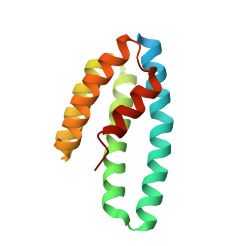
    2N8I, 2N8W, 5E6G

  • PubMed Abstract: 

    Natural recombination combines pieces of preexisting proteins to create new tertiary structures and functions. We describe a computational protocol, called SEWING, which is inspired by this process and builds new proteins from connected or disconnected pieces of existing structures. Helical proteins designed with SEWING contain structural features absent from other de novo designed proteins and, in some cases, remain folded at more than 100°C. High-resolution structures of the designed proteins CA01 and DA05R1 were solved by x-ray crystallography (2.2 angstrom resolution) and nuclear magnetic resonance, respectively, and there was excellent agreement with the design models. This method provides a new strategy to rapidly create large numbers of diverse and designable protein scaffolds.


  • Organizational Affiliation
    • Program in Bioinformatics and Computational Biology, University of North Carolina at Chapel Hill, Chapel Hill, NC 27599, USA.

Macromolecule Content 

  • Total Structure Weight: 31.44 kDa 
  • Atom Count: 1,980 
  • Modeled Residue Count: 227 
  • Deposited Residue Count: 272 
  • Unique protein chains: 1

Macromolecules

Find similar proteins by:|  3D Structure
Entity ID: 1
MoleculeChains  Sequence LengthOrganismDetailsImage
De novo designed protein CA01
A, B
136synthetic constructMutation(s): 0 

Experimental Data & Validation

Experimental Data

  • Method: X-RAY DIFFRACTION
  • Resolution: 2.09 Å
  • R-Value Free:  0.234 (Depositor), 0.241 (DCC) 
  • R-Value Work:  0.200 (Depositor), 0.208 (DCC) 
  • R-Value Observed: 0.202 (Depositor) 
Space Group: H 3
Unit Cell:
Length ( Å )Angle ( ˚ )
a = 116.537α = 90
b = 116.537β = 90
c = 57.215γ = 120
Software Package:
Software NamePurpose
PHENIXrefinement
SCALAdata scaling
PHASERphasing

Structure Validation

View Full Validation Report


Entry History 

& Funding Information

Deposition Data


Funding OrganizationLocationGrant Number
National Institutes of Health/National Institute of General Medical Sciences (NIH/NIGMS)United StatesR01-GM073960

Revision History  (Full details and data files)

  • Version 1.0: 2016-05-18
    Type: Initial release
  • Version 1.1: 2017-09-20
    Changes: Author supporting evidence, Database references, Derived calculations
  • Version 1.2: 2017-11-01
    Changes: Author supporting evidence
  • Version 1.3: 2019-12-25
    Changes: Author supporting evidence
  • Version 1.4: 2024-03-06
    Changes: Data collection, Database references