


Crystal structures of uPARAP, a member of mannose receptor family
Yuan, C., Huang, M.To be published.
Experimental Data Snapshot
Starting Models: experimental
View more details
Entity ID: 1 | |||||
|---|---|---|---|---|---|
| Molecule | Chains | Sequence Length | Organism | Details | Image |
| C-type mannose receptor 2 | 492 | Homo sapiens | Mutation(s): 1 Gene Names: MRC2, CLEC13E, ENDO180, KIAA0709, UPARAP | 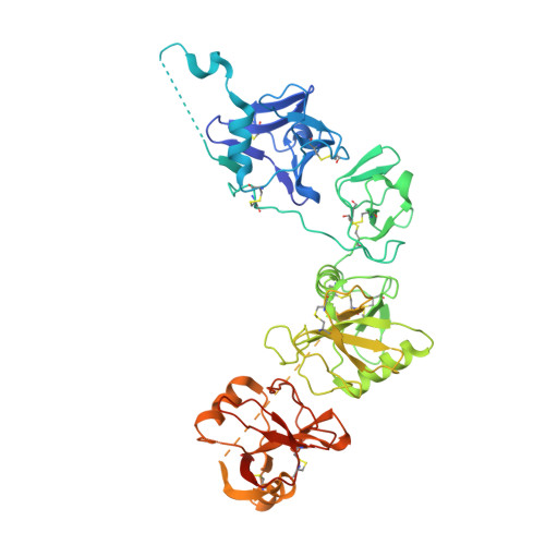 | |
UniProt & NIH Common Fund Data Resources | |||||
PHAROS: Q9UBG0 GTEx: ENSG00000011028 | |||||
Entity Groups | |||||
| Sequence Clusters | 30% Identity50% Identity70% Identity90% Identity95% Identity100% Identity | ||||
| UniProt Group | Q9UBG0 | ||||
Glycosylation | |||||
| Glycosylation Sites: 1 | Go to GlyGen: Q9UBG0-1 | ||||
Sequence AnnotationsExpand | |||||
Reference Sequence | |||||
| Ligands 2 Unique | |||||
|---|---|---|---|---|---|
| ID | Chains | Name / Formula / InChI Key | 2D Diagram | 3D Interactions | |
| NAG Download:Ideal Coordinates CCD File | F [auth A], J [auth B] | 2-acetamido-2-deoxy-beta-D-glucopyranose C8 H15 N O6 OVRNDRQMDRJTHS-FMDGEEDCSA-N |  | ||
| CA Download:Ideal Coordinates CCD File | C [auth A] D [auth A] E [auth A] G [auth B] H [auth B] | CALCIUM ION Ca BHPQYMZQTOCNFJ-UHFFFAOYSA-N |  | ||
| Length ( Å ) | Angle ( ˚ ) |
|---|---|
| a = 74.27 | α = 90 |
| b = 102.7 | β = 94.54 |
| c = 87.76 | γ = 90 |
| Software Name | Purpose |
|---|---|
| PHENIX | refinement |
| xia2 | data scaling |
| PDB_EXTRACT | data extraction |
| xia2 | data reduction |
| PHENIX | phasing |
| Funding Organization | Location | Grant Number |
|---|---|---|
| Natural Science Foundation Of China | China | 31570745 |


