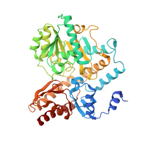
Crystal structure of the ornithine aminotransferase from Toxoplasma gondii ME49 in a complex with (S)-4-amino-5-fluoropentanoic acid
Filippova, E.V., Minasov, G., Flores, K., Le, H.V., Silverman, R.B., McLeod, R.L., Anderson, W.F., Center for Structural Genomics of Infectious Diseases (CSGID)To be published.