5E1I | pdb_00005e1i

Crystal structure of Mycobacterium tuberculosis L,D-transpeptidase 2 with carbapenem drug T210


Experimental Data Snapshot

  • Method: X-RAY DIFFRACTION
  • Resolution: 2.00 Å
  • R-Value Free: 
    0.225 (Depositor), 0.195 (DCC) 
  • R-Value Work: 
    0.191 (Depositor), 0.195 (DCC) 
  • R-Value Observed: 
    0.193 (Depositor) 

Starting Model: experimental
View more details

wwPDB Validation 3D Report Full Report

Validation slider image for 5E1I

Ligand Structure Quality Assessment 


This is version 1.6 of the entry. See complete history

Literature

Non-classical transpeptidases yield insight into new antibacterials.

Kumar, P.Kaushik, A.Lloyd, E.P.Li, S.G.Mattoo, R.Ammerman, N.C.Bell, D.T.Perryman, A.L.Zandi, T.A.Ekins, S.Ginell, S.L.Townsend, C.A.Freundlich, J.S.Lamichhane, G.

(2017) Nat Chem Biol 13: 54-61

  • DOI: https://doi.org/10.1038/nchembio.2237
  • Primary Citation Related Structures: 
    5DU7, 5DUJ, 5DVP, 5DZJ, 5DZP, 5E1G, 5E1I, 5E51, 5E5L, 5K69

  • PubMed Abstract: 

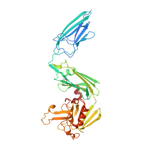
    Bacterial survival requires an intact peptidoglycan layer, a three-dimensional exoskeleton that encapsulates the cytoplasmic membrane. Historically, the final steps of peptidoglycan synthesis are known to be carried out by D,D-transpeptidases, enzymes that are inhibited by the β-lactams, which constitute >50% of all antibacterials in clinical use. Here, we show that the carbapenem subclass of β-lactams are distinctly effective not only because they inhibit D,D-transpeptidases and are poor substrates for β-lactamases, but primarily because they also inhibit non-classical transpeptidases, namely the L,D-transpeptidases, which generate the majority of linkages in the peptidoglycan of mycobacteria. We have characterized the molecular mechanisms responsible for inhibition of L,D-transpeptidases of Mycobacterium tuberculosis and a range of bacteria including ESKAPE pathogens, and used this information to design, synthesize and test simplified carbapenems with potent antibacterial activity.


  • Organizational Affiliation
    • Department of Medicine, Johns Hopkins University, Baltimore, Maryland, USA.

Macromolecule Content 

  • Total Structure Weight: 79.44 kDa 
  • Atom Count: 5,623 
  • Modeled Residue Count: 701 
  • Deposited Residue Count: 740 
  • Unique protein chains: 1

Macromolecules

Find similar proteins by:|  3D Structure
Entity ID: 1
MoleculeChains  Sequence LengthOrganismDetailsImage
L,D-transpeptidase 2
A, B
370Mycobacterium tuberculosis H37RvMutation(s): 0 
Gene Names: ldtBlppSRv2518cRVBD_2518cP425_02624
EC: 2.3.2
UniProt
Find proteins for I6Y9J2 (Mycobacterium tuberculosis (strain ATCC 25618 / H37Rv))
Explore I6Y9J2 
Go to UniProtKB:  I6Y9J2
Entity Groups
Sequence Clusters30% Identity50% Identity70% Identity90% Identity95% Identity100% Identity
UniProt GroupI6Y9J2
Sequence Annotations
Expand
Reference Sequence

Small Molecules

Ligands 3 Unique
IDChains Name / Formula / InChI Key2D Diagram3D Interactions
6B8

Query on 6B8


Download:Ideal Coordinates CCD File
D [auth A](2S,3R,4R)-2-[(2S,3R)-3-hydroxy-1-oxobutan-2-yl]-3-methyl-4-(methylsulfanyl)-3,4-dihydro-2H-pyrrole-5-carboxylic acid
C11 H17 N O4 S
ASUOZXIKIDLJND-VRRGKTLJSA-N
GOL

Query on GOL


Download:Ideal Coordinates CCD File
E [auth A]GLYCEROL
C3 H8 O3
PEDCQBHIVMGVHV-UHFFFAOYSA-N
NA

Query on NA


Download:Ideal Coordinates CCD File
C [auth A]SODIUM ION
Na
FKNQFGJONOIPTF-UHFFFAOYSA-N

Experimental Data & Validation

Experimental Data

  • Method: X-RAY DIFFRACTION
  • Resolution: 2.00 Å
  • R-Value Free:  0.225 (Depositor), 0.195 (DCC) 
  • R-Value Work:  0.191 (Depositor), 0.195 (DCC) 
  • R-Value Observed: 0.193 (Depositor) 
Space Group: P 1 21 1
Unit Cell:
Length ( Å )Angle ( ˚ )
a = 61.183α = 90
b = 93.957β = 93.04
c = 75.391γ = 90
Software Package:
Software NamePurpose
PHENIXrefinement
HKL-2000data scaling
PDB_EXTRACTdata extraction
HKL-3000data reduction
PHASERphasing

Structure Validation

View Full Validation Report


Ligand Structure Quality Assessment 


Entry History 

& Funding Information

Deposition Data


Funding OrganizationLocationGrant Number
National Institutes of Health/Office of the DirectorUnited States1DP2OD008459-01

Revision History  (Full details and data files)

  • Version 1.0: 2016-10-12
    Type: Initial release
  • Version 1.1: 2016-11-09
    Changes: Database references
  • Version 1.2: 2016-11-23
    Changes: Database references
  • Version 1.3: 2016-12-28
    Changes: Database references
  • Version 1.4: 2017-09-20
    Changes: Author supporting evidence
  • Version 1.5: 2023-09-27
    Changes: Data collection, Database references, Derived calculations, Refinement description
  • Version 1.6: 2024-10-09
    Changes: Structure summary