5E0I | pdb_00005e0i

Crystal structure of the HBV capsid Y132A mutant (VCID 8772) in complex with NVR10-001E2 at 1.95A resolution


Experimental Data Snapshot

  • Method: X-RAY DIFFRACTION
  • Resolution: 1.95 Å
  • R-Value Free: 
    0.252 (Depositor), 0.262 (DCC) 
  • R-Value Work: 
    0.219 (Depositor), 0.232 (DCC) 
  • R-Value Observed: 
    0.221 (Depositor) 

Starting Model: experimental
View more details

wwPDB Validation 3D Report Full Report

Validation slider image for 5E0I

Ligand Structure Quality Assessment 


This is version 1.3 of the entry. See complete history

Literature

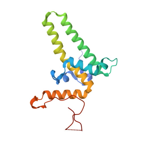
High-resolution crystal structure of a hepatitis B virus replication inhibitor bound to the viral core protein.

Klumpp, K.Lam, A.M.Lukacs, C.Vogel, R.Ren, S.Espiritu, C.Baydo, R.Atkins, K.Abendroth, J.Liao, G.Efimov, A.Hartman, G.Flores, O.A.

(2015) Proc Natl Acad Sci U S A 112: 15196-15201

  • DOI: https://doi.org/10.1073/pnas.1513803112
  • Primary Citation Related Structures: 
    5E0I

  • PubMed Abstract: 

    The hepatitis B virus (HBV) core protein is essential for HBV replication and an important target for antiviral drug discovery. We report the first, to our knowledge, high-resolution crystal structure of an antiviral compound bound to the HBV core protein. The compound NVR-010-001-E2 can induce assembly of the HBV core wild-type and Y132A mutant proteins and thermostabilize the proteins with a Tm increase of more than 10 °C. NVR-010-001-E2 binds at the dimer-dimer interface of the core proteins, forms a new interaction surface promoting protein-protein interaction, induces protein assembly, and increases stability. The impact of naturally occurring core protein mutations on antiviral activity correlates with NVR-010-001-E2 binding interactions determined by crystallography. The crystal structure provides understanding of a drug efficacy mechanism related to the induction and stabilization of protein-protein interactions and enables structure-guided design to improve antiviral potency and drug-like properties.


  • Organizational Affiliation
    • Novira Therapeutics, Doylestown, PA 18902; kklumpp@noviratherapeutics.com.

Macromolecule Content 

  • Total Structure Weight: 109.09 kDa 
  • Atom Count: 7,532 
  • Modeled Residue Count: 880 
  • Deposited Residue Count: 942 
  • Unique protein chains: 1

Macromolecules

Find similar proteins by:|  3D Structure
Entity ID: 1
MoleculeChains  Sequence LengthOrganismDetailsImage
Capsid protein
A, B, C, D, E
A, B, C, D, E, F
157Hepatitis B virus subtype adywMutation(s): 1 
UniProt
Find proteins for P03147 (Hepatitis B virus genotype D subtype adw (isolate United Kingdom/adyw/1979))
Explore P03147 
Go to UniProtKB:  P03147
Entity Groups
Sequence Clusters30% Identity50% Identity70% Identity90% Identity95% Identity100% Identity
UniProt GroupP03147
Sequence Annotations
Expand
Reference Sequence

Experimental Data & Validation

Experimental Data

  • Method: X-RAY DIFFRACTION
  • Resolution: 1.95 Å
  • R-Value Free:  0.252 (Depositor), 0.262 (DCC) 
  • R-Value Work:  0.219 (Depositor), 0.232 (DCC) 
  • R-Value Observed: 0.221 (Depositor) 
Space Group: C 1 2 1
Unit Cell:
Length ( Å )Angle ( ˚ )
a = 152.6α = 90
b = 88.17β = 103.47
c = 102.25γ = 90
Software Package:
Software NamePurpose
XDSdata reduction
XSCALEdata scaling
Cootmodel building
PHASERphasing
REFMACrefinement

Structure Validation

View Full Validation Report


Ligand Structure Quality Assessment 


Entry History 

Deposition Data

Revision History  (Full details and data files)

  • Version 1.0: 2015-12-09
    Type: Initial release
  • Version 1.1: 2015-12-16
    Changes: Database references
  • Version 1.2: 2023-09-27
    Changes: Data collection, Database references, Derived calculations, Refinement description
  • Version 1.3: 2024-11-13
    Changes: Structure summary