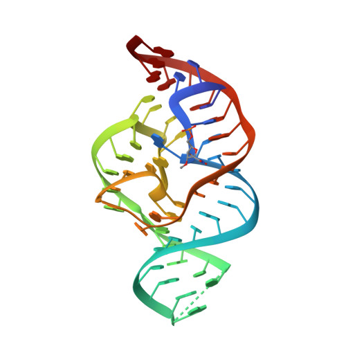
5DUN | pdb_00005dun

The crystal structure of OMe substituted twister ribozyme


Experimental Data Snapshot

  • Method: X-RAY DIFFRACTION
  • Resolution: 2.64 Å
  • R-Value Free: 
    0.252 (Depositor), 0.259 (DCC) 
  • R-Value Work: 
    0.194 (Depositor), 0.204 (DCC) 
  • R-Value Observed: 
    0.197 (Depositor) 

Starting Model: experimental
View more details

wwPDB Validation 3D Report Full Report

Validation slider image for 5DUN

This is version 1.2 of the entry. See complete history. 

Literature

A Mini-Twister Variant and Impact of Residues/Cations on the Phosphodiester Cleavage of this Ribozyme Class.

Kosutic, M., Neuner, S., Ren, A., Flur, S., Wunderlich, C., Mairhofer, E., Vusurovic, N., Seikowski, J., Breuker, K., Hobartner, C., Patel, D.J., Kreutz, C., Micura, R.

(2015) Angew Chem Int Ed Engl 54: 15128-15133

  • DOI: https://doi.org/10.1002/anie.201506601
  • Primary Citation Related Structures: 
    5DUN

  • PubMed Abstract: 

    Nucleolytic ribozymes catalyze site-specific cleavage of their phosphodiester backbones. A minimal version of the twister ribozyme is reported that lacks the phylogenetically conserved stem P1 while retaining wild-type activity. Atomic mutagenesis revealed that nitrogen atoms N1 and N3 of the adenine-6 at the cleavage site are indispensable for cleavage. By NMR spectroscopy, a pKa value of 5.1 was determined for a (13) C2-labeled adenine at this position in the twister ribozyme, which is significantly shifted compared to the pKa of the same adenine in the substrate alone. This finding pinpoints at a potential role for adenine-6 in the catalytic mechanism besides the previously identified invariant guanine-48 and a Mg(2+) ion, both of which are directly coordinated to the non-bridging oxygen atoms of the scissile phosphate; for the latter, additional evidence stems from the observation that Mn(2+) or Cd(2+) accelerated cleavage of phosphorothioate substrates. The relevance of this metal ion binding site is further emphasized by a new 2.6 Å X-ray structure of a 2'-OCH3 -U5 modified twister ribozyme.


  • Organizational Affiliation: 
    • Institute of Organic Chemistry and Center for Molecular Biosciences CMBI, Leopold-Franzens University, Innrain 80-82, 6020 Innsbruck (Austria).

Macromolecule Content 

  • Total Structure Weight: 17.64 kDa 
  • Atom Count: 1,179 
  • Modeled Residue Count: 54 
  • Deposited Residue Count: 54 
  • Unique nucleic acid chains: 1

Macromolecules

Find similar nucleic acids by:  (by identity cutoff) 
Entity ID: 1
MoleculeChains LengthOrganismImage
RNA (54-MER)54synthetic construct
Sequence Annotations
Expand
Reference Sequence

Small Molecules

Ligands 2 Unique
IDChains Name / Formula / InChI Key2D Diagram3D Interactions
K

Query on K


Download:Ideal Coordinates CCD File
J [auth A]POTASSIUM ION
K
NPYPAHLBTDXSSS-UHFFFAOYSA-N
MG

Query on MG


Download:Ideal Coordinates CCD File
B [auth A]
C [auth A]
D [auth A]
E [auth A]
F [auth A]
B [auth A],
C [auth A],
D [auth A],
E [auth A],
F [auth A],
G [auth A],
H [auth A],
I [auth A]
MAGNESIUM ION
Mg
JLVVSXFLKOJNIY-UHFFFAOYSA-N

Experimental Data & Validation

Experimental Data

  • Method: X-RAY DIFFRACTION
  • Resolution: 2.64 Å
  • R-Value Free:  0.252 (Depositor), 0.259 (DCC) 
  • R-Value Work:  0.194 (Depositor), 0.204 (DCC) 
  • R-Value Observed: 0.197 (Depositor) 
Space Group: P 43 21 2
Unit Cell:
Length ( Å )Angle ( ˚ )
a = 46.819α = 90
b = 46.819β = 90
c = 143.751γ = 90
Software Package:
Software NamePurpose
PHENIXrefinement
XDSdata reduction
XDSdata scaling
PHASERphasing

Structure Validation

View Full Validation Report


Entry History 

Deposition Data

Revision History  (Full details and data files)

  • Version 1.0: 2015-10-28
    Type: Initial release
  • Version 1.1: 2016-01-27
    Changes: Database references
  • Version 1.2: 2023-09-27
    Changes: Data collection, Database references, Derived calculations, Refinement description