5DS5 | pdb_00005ds5

Crystal structure the Escherichia coli Cas1-Cas2 complex bound to protospacer DNA and Mg


Experimental Data Snapshot

  • Method: X-RAY DIFFRACTION
  • Resolution: 2.95 Å
  • R-Value Free: 
    0.256 (Depositor), 0.255 (DCC) 
  • R-Value Work: 
    0.234 (Depositor), 0.236 (DCC) 
  • R-Value Observed: 
    0.235 (Depositor) 

Starting Model: experimental
View more details

wwPDB Validation 3D Report Full Report

Validation slider image for 5DS5

This is version 1.5 of the entry. See complete history

Literature

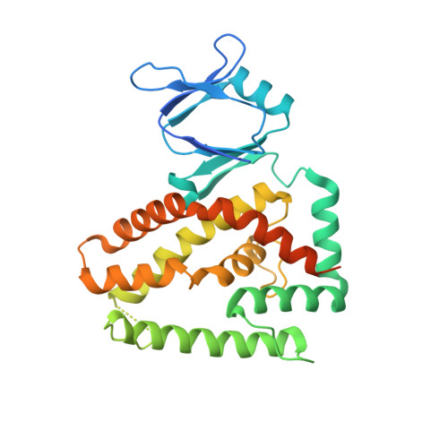
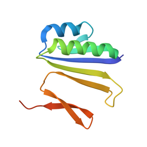
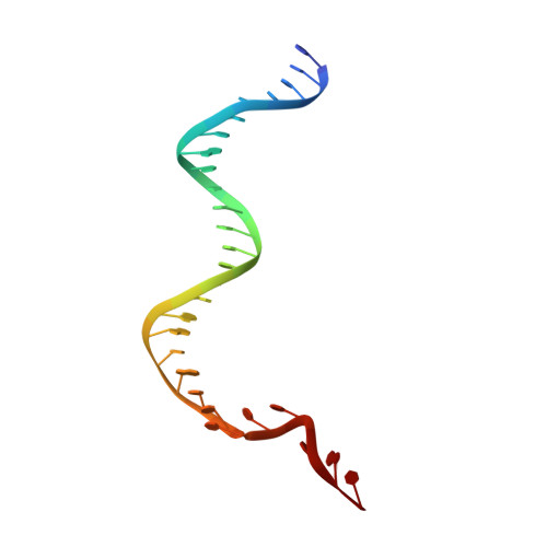
Foreign DNA capture during CRISPR-Cas adaptive immunity.

Nunez, J.K.Harrington, L.B.Kranzusch, P.J.Engelman, A.N.Doudna, J.A.

(2015) Nature 527: 535-538

  • DOI: https://doi.org/10.1038/nature15760
  • Primary Citation Related Structures: 
    5DS4, 5DS5, 5DS6

  • PubMed Abstract: 

    Bacteria and archaea generate adaptive immunity against phages and plasmids by integrating foreign DNA of specific 30-40-base-pair lengths into clustered regularly interspaced short palindromic repeat (CRISPR) loci as spacer segments. The universally conserved Cas1-Cas2 integrase complex catalyses spacer acquisition using a direct nucleophilic integration mechanism similar to retroviral integrases and transposases. How the Cas1-Cas2 complex selects foreign DNA substrates for integration remains unknown. Here we present X-ray crystal structures of the Escherichia coli Cas1-Cas2 complex bound to cognate 33-nucleotide protospacer DNA substrates. The protein complex creates a curved binding surface spanning the length of the DNA and splays the ends of the protospacer to allow each terminal nucleophilic 3'-OH to enter a channel leading into the Cas1 active sites. Phosphodiester backbone interactions between the protospacer and the proteins explain the sequence-nonspecific substrate selection observed in vivo. Our results uncover the structural basis for foreign DNA capture and the mechanism by which Cas1-Cas2 functions as a molecular ruler to dictate the sequence architecture of CRISPR loci.


  • Organizational Affiliation
    • Department of Molecular and Cell Biology, University of California, Berkeley, Berkeley, California 94720, USA.

Macromolecule Content 

  • Total Structure Weight: 174 kDa 
  • Atom Count: 10,719 
  • Modeled Residue Count: 1,298 
  • Deposited Residue Count: 1,488 
  • Unique protein chains: 2
  • Unique nucleic acid chains: 2

Macromolecules


Find similar proteins by:|  3D Structure
Entity ID: 1
MoleculeChains  Sequence LengthOrganismDetailsImage
CRISPR-associated endonuclease Cas1
A, B, C, D
306Escherichia coli K-12Mutation(s): 0 
Gene Names: ygbTcas1b2755JW2725
EC: 3.1
UniProt
Find proteins for Q46896 (Escherichia coli (strain K12))
Explore Q46896 
Go to UniProtKB:  Q46896
Entity Groups
Sequence Clusters30% Identity50% Identity70% Identity90% Identity95% Identity100% Identity
UniProt GroupQ46896
Sequence Annotations
Expand
Reference Sequence
Find similar proteins by:|  3D Structure
Entity ID: 2
MoleculeChains  Sequence LengthOrganismDetailsImage
CRISPR-associated endoribonuclease Cas2
E, F
104Escherichia coli K-12Mutation(s): 0 
Gene Names: ygbFcas2b2754JW5438
EC: 3.1
UniProt
Find proteins for P45956 (Escherichia coli (strain K12))
Explore P45956 
Go to UniProtKB:  P45956
Entity Groups
Sequence Clusters30% Identity50% Identity70% Identity90% Identity95% Identity100% Identity
UniProt GroupP45956
Sequence Annotations
Expand
Reference Sequence
Find similar nucleic acids by:  (by identity cutoff) 
Entity ID: 3
MoleculeChains LengthOrganismImage
DNA (28-MER)28Inovirus M13
Sequence Annotations
Expand
Reference Sequence
Find similar nucleic acids by:  (by identity cutoff) 
Entity ID: 4
MoleculeChains LengthOrganismImage
DNA (28-MER)28Inovirus M13
Sequence Annotations
Expand
Reference Sequence

Experimental Data & Validation

Experimental Data

  • Method: X-RAY DIFFRACTION
  • Resolution: 2.95 Å
  • R-Value Free:  0.256 (Depositor), 0.255 (DCC) 
  • R-Value Work:  0.234 (Depositor), 0.236 (DCC) 
  • R-Value Observed: 0.235 (Depositor) 
Space Group: P 21 21 21
Unit Cell:
Length ( Å )Angle ( ˚ )
a = 75.656α = 90
b = 165.928β = 90
c = 167.258γ = 90
Software Package:
Software NamePurpose
PHENIXrefinement
XDSdata reduction
Aimlessdata scaling
PHENIXphasing

Structure Validation

View Full Validation Report


Entry History 

& Funding Information

Deposition Data


Funding OrganizationLocationGrant Number
Howard Hughes Medical Institute (HHMI)United States--

Revision History  (Full details and data files)

  • Version 1.0: 2015-10-28
    Type: Initial release
  • Version 1.1: 2015-11-11
    Changes: Database references
  • Version 1.2: 2015-12-09
    Changes: Database references
  • Version 1.3: 2018-01-17
    Changes: Author supporting evidence, Database references, Derived calculations
  • Version 1.4: 2019-11-20
    Changes: Author supporting evidence
  • Version 1.5: 2023-09-27
    Changes: Data collection, Database references, Derived calculations, Refinement description