5DLT | pdb_00005dlt

Crystal structure of Autotaxin (ENPP2) with 7-alpha-hydroxycholesterol


Experimental Data Snapshot

  • Method: X-RAY DIFFRACTION
  • Resolution: 1.60 Å
  • R-Value Free: 
    0.196 (Depositor), 0.203 (DCC) 
  • R-Value Work: 
    0.171 (Depositor) 
  • R-Value Observed: 
    0.172 (Depositor) 

Starting Model: experimental
View more details

wwPDB Validation 3D Report Full Report

Validation slider image for 5DLT

Ligand Structure Quality Assessment 


This is version 2.2 of the entry. See complete history

Literature

Steroid binding to Autotaxin links bile salts and lysophosphatidic acid signalling.

Keune, W.J.Hausmann, J.Bolier, R.Tolenaars, D.Kremer, A.Heidebrecht, T.Joosten, R.P.Sunkara, M.Morris, A.J.Matas-Rico, E.Moolenaar, W.H.Oude Elferink, R.P.Perrakis, A.

(2016) Nat Commun 7: 11248-11248

  • DOI: https://doi.org/10.1038/ncomms11248
  • Primary Citation Related Structures: 
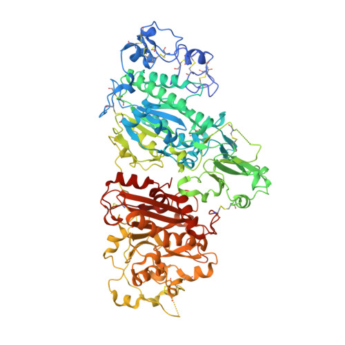
    5DLT, 5DLV, 5DLW

  • PubMed Abstract: 

    Autotaxin (ATX) generates the lipid mediator lysophosphatidic acid (LPA). ATX-LPA signalling is involved in multiple biological and pathophysiological processes, including vasculogenesis, fibrosis, cholestatic pruritus and tumour progression. ATX has a tripartite active site, combining a hydrophilic groove, a hydrophobic lipid-binding pocket and a tunnel of unclear function. We present crystal structures of rat ATX bound to 7α-hydroxycholesterol and the bile salt tauroursodeoxycholate (TUDCA), showing how the tunnel selectively binds steroids. A structure of ATX simultaneously harbouring TUDCA in the tunnel and LPA in the pocket, together with kinetic analysis, reveals that bile salts act as partial non-competitive inhibitors of ATX, thereby attenuating LPA receptor activation. This unexpected interplay between ATX-LPA signalling and select steroids, notably natural bile salts, provides a molecular basis for the emerging association of ATX with disorders associated with increased circulating levels of bile salts. Furthermore, our findings suggest potential clinical implications in the use of steroid drugs.


  • Organizational Affiliation
    • Division of Biochemistry, Netherlands Cancer Institute, 1066 CX Amsterdam, The Netherlands.

Macromolecule Content 

  • Total Structure Weight: 98.88 kDa 
  • Atom Count: 7,189 
  • Modeled Residue Count: 796 
  • Deposited Residue Count: 827 
  • Unique protein chains: 1

Macromolecules

Find similar proteins by:|  3D Structure
Entity ID: 1
MoleculeChains  Sequence LengthOrganismDetailsImage
Ectonucleotide pyrophosphatase/phosphodiesterase family member 2827Rattus norvegicusMutation(s): 2 
Gene Names: Enpp2AtxNpps2
EC: 3.1.4.39 (PDB Primary Data), 3.1.4.4 (UniProt)
UniProt
Find proteins for Q64610 (Rattus norvegicus)
Explore Q64610 
Go to UniProtKB:  Q64610
Entity Groups
Sequence Clusters30% Identity50% Identity70% Identity90% Identity95% Identity100% Identity
UniProt GroupQ64610
Glycosylation
Glycosylation Sites: 1Go to GlyGen: Q64610-1
Sequence Annotations
Expand
Reference Sequence

Oligosaccharides

Help  
Entity ID: 2
MoleculeChains Length2D Diagram GlycosylationD Interactions
beta-D-mannopyranose-(1-4)-2-acetamido-2-deoxy-beta-D-glucopyranose-(1-4)-2-acetamido-2-deoxy-beta-D-glucopyranose
B
3N-Glycosylation
Glycosylation Resources
GlyTouCan: G15407YE
GlyCosmos: G15407YE
GlyGen: G15407YE

Small Molecules

Ligands 7 Unique
IDChains Name / Formula / InChI Key2D Diagram3D Interactions
5JK

Query on 5JK


Download:Ideal Coordinates CCD File
E [auth A]7alpha-hydroxycholesterol
C27 H46 O2
OYXZMSRRJOYLLO-RVOWOUOISA-N
IOD

Query on IOD


Download:Ideal Coordinates CCD File
G [auth A]
H [auth A]
I [auth A]
J [auth A]
K [auth A]
G [auth A],
H [auth A],
I [auth A],
J [auth A],
K [auth A],
L [auth A],
M [auth A],
N [auth A],
O [auth A],
P [auth A]
IODIDE ION
I
XMBWDFGMSWQBCA-UHFFFAOYSA-M
GOL

Query on GOL


Download:Ideal Coordinates CCD File
CA [auth A]
DA [auth A]
EA [auth A]
FA [auth A]
GA [auth A]
CA [auth A],
DA [auth A],
EA [auth A],
FA [auth A],
GA [auth A],
HA [auth A],
IA [auth A]
GLYCEROL
C3 H8 O3
PEDCQBHIVMGVHV-UHFFFAOYSA-N
ZN

Query on ZN


Download:Ideal Coordinates CCD File
C [auth A],
D [auth A]
ZINC ION
Zn
PTFCDOFLOPIGGS-UHFFFAOYSA-N
SCN

Query on SCN


Download:Ideal Coordinates CCD File
JA [auth A]
Q [auth A]
R [auth A]
S [auth A]
T [auth A]
JA [auth A],
Q [auth A],
R [auth A],
S [auth A],
T [auth A],
U [auth A],
V [auth A],
W [auth A],
X [auth A],
Y [auth A],
Z [auth A]
THIOCYANATE ION
C N S
ZMZDMBWJUHKJPS-UHFFFAOYSA-M
CA

Query on CA


Download:Ideal Coordinates CCD File
F [auth A]CALCIUM ION
Ca
BHPQYMZQTOCNFJ-UHFFFAOYSA-N
NA

Query on NA


Download:Ideal Coordinates CCD File
AA [auth A],
BA [auth A]
SODIUM ION
Na
FKNQFGJONOIPTF-UHFFFAOYSA-N
Modified Residues  1 Unique
IDChains TypeFormula2D DiagramParent
TPO
Query on TPO
A
L-PEPTIDE LINKINGC4 H10 N O6 PTHR

Experimental Data & Validation

Experimental Data

  • Method: X-RAY DIFFRACTION
  • Resolution: 1.60 Å
  • R-Value Free:  0.196 (Depositor), 0.203 (DCC) 
  • R-Value Work:  0.171 (Depositor) 
  • R-Value Observed: 0.172 (Depositor) 
Space Group: P 1
Unit Cell:
Length ( Å )Angle ( ˚ )
a = 53.671α = 98.72
b = 63.482β = 105.8
c = 70.715γ = 99.97
Software Package:
Software NamePurpose
REFMACrefinement
PDB_EXTRACTdata extraction
Aimlessdata scaling
XDSdata reduction
PHASERphasing
Cootmodel building

Structure Validation

View Full Validation Report


Ligand Structure Quality Assessment 


Entry History 

& Funding Information

Deposition Data


Funding OrganizationLocationGrant Number
NWONetherlands700.10.354
NWONetherlands723.013.003

Revision History  (Full details and data files)

  • Version 1.0: 2016-04-13
    Type: Initial release
  • Version 1.1: 2016-04-27
    Changes: Database references
  • Version 2.0: 2020-07-29
    Type: Remediation
    Reason: Carbohydrate remediation
    Changes: Atomic model, Data collection, Derived calculations, Structure summary
  • Version 2.1: 2024-01-10
    Changes: Data collection, Database references, Refinement description, Structure summary
  • Version 2.2: 2024-11-13
    Changes: Structure summary