5DKT | pdb_00005dkt

N-terminal His tagged apPOL exonuclease mutant


Experimental Data Snapshot

  • Method: X-RAY DIFFRACTION
  • Resolution: 2.90 Å
  • R-Value Free: 
    0.299 (Depositor), 0.301 (DCC) 
  • R-Value Work: 
    0.265 (Depositor), 0.268 (DCC) 
  • R-Value Observed: 
    0.268 (Depositor) 

Starting Model: experimental
View more details

wwPDB Validation 3D Report Full Report

Validation slider image for 5DKT

This is version 1.3 of the entry. See complete history

Literature

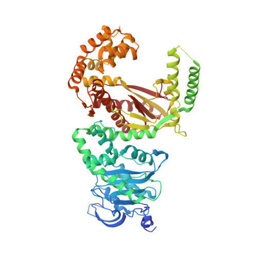
Crystal Structure of the Apicoplast DNA Polymerase from Plasmodium falciparum: The First Look at a Plastidic A-Family DNA Polymerase.

Milton, M.E.Choe, J.Y.Honzatko, R.B.Nelson, S.W.

(2016) J Mol Biology 428: 3920-3934

  • DOI: https://doi.org/10.1016/j.jmb.2016.07.016
  • Primary Citation Related Structures: 
    5DKT, 5DKU

  • PubMed Abstract: 

    Plasmodium falciparum, the primary cause of malaria, contains a non-photosynthetic plastid called the apicoplast. The apicoplast exists in most members of the phylum Apicomplexa and has its own genome along with organelle-specific enzymes for its replication. The only DNA polymerase found in the apicoplast (apPOL) was putatively acquired through horizontal gene transfer from a bacteriophage and is classified as an atypical A-family polymerase. Here, we present its crystal structure at a resolution of 2.9Å. P. falciparum apPOL, the first structural representative of a plastidic A-family polymerase, diverges from typical A-family members in two of three previously identified signature motifs and in a region not implicated by sequence. Moreover, apPOL has an additional N-terminal subdomain, the absence of which severely diminishes its 3' to 5' exonuclease activity. A compound known to be toxic to Plasmodium is a potent inhibitor of apPOL, suggesting that apPOL is a viable drug target. The structure provides new insights into the structural diversity of A-family polymerases and may facilitate structurally guided antimalarial drug design.


  • Organizational Affiliation
    • Department of Biochemistry, Biophysics, and Molecular Biology, Iowa State University, Ames, IA 50011, USA.

Macromolecule Content 

  • Total Structure Weight: 77.06 kDa 
  • Atom Count: 4,909 
  • Modeled Residue Count: 577 
  • Deposited Residue Count: 648 
  • Unique protein chains: 1

Macromolecules

Find similar proteins by:|  3D Structure
Entity ID: 1
MoleculeChains  Sequence LengthOrganismDetailsImage
Prex DNA polymerase648Plasmodium falciparum 3D7Mutation(s): 2 
Gene Names: POM1PF14_0112
EC: 2.7.7.7 (PDB Primary Data), 2.7.7.102 (UniProt), 5.6.2.3 (UniProt), 3.1.11 (UniProt)
UniProt
Find proteins for Q8ILY1 (Plasmodium falciparum (isolate 3D7))
Explore Q8ILY1 
Go to UniProtKB:  Q8ILY1
Entity Groups
Sequence Clusters30% Identity50% Identity70% Identity90% Identity95% Identity100% Identity
UniProt GroupQ8ILY1
Sequence Annotations
Expand
Reference Sequence

Small Molecules

Ligands 2 Unique
IDChains Name / Formula / InChI Key2D Diagram3D Interactions
SO4

Query on SO4


Download:Ideal Coordinates CCD File
B [auth A]
C [auth A]
D [auth A]
E [auth A]
F [auth A]
B [auth A],
C [auth A],
D [auth A],
E [auth A],
F [auth A],
G [auth A],
H [auth A],
I [auth A],
J [auth A]
SULFATE ION
O4 S
QAOWNCQODCNURD-UHFFFAOYSA-L
NA

Query on NA


Download:Ideal Coordinates CCD File
K [auth A]SODIUM ION
Na
FKNQFGJONOIPTF-UHFFFAOYSA-N

Experimental Data & Validation

Experimental Data

  • Method: X-RAY DIFFRACTION
  • Resolution: 2.90 Å
  • R-Value Free:  0.299 (Depositor), 0.301 (DCC) 
  • R-Value Work:  0.265 (Depositor), 0.268 (DCC) 
  • R-Value Observed: 0.268 (Depositor) 
Space Group: P 65 2 2
Unit Cell:
Length ( Å )Angle ( ˚ )
a = 141.775α = 90
b = 141.775β = 90
c = 148.759γ = 120
Software Package:
Software NamePurpose
PHENIXrefinement
HKL-3000data reduction
HKL-3000data scaling
MR-Rosettaphasing

Structure Validation

View Full Validation Report


Entry History 

& Funding Information

Deposition Data


Funding OrganizationLocationGrant Number
Iowa State University College of Liberal Arts and SciencesUnited States--

Revision History  (Full details and data files)

  • Version 1.0: 2016-08-10
    Type: Initial release
  • Version 1.1: 2016-08-17
    Changes: Database references
  • Version 1.2: 2016-10-19
    Changes: Database references
  • Version 1.3: 2023-09-27
    Changes: Data collection, Database references, Derived calculations, Refinement description