5DJC | pdb_00005djc

Fc Heterodimer Design 8.1 L368V/Y407A + T366V/K409F


Experimental Data Snapshot

  • Method: X-RAY DIFFRACTION
  • Resolution: 2.10 Å
  • R-Value Free: 
    0.285 (Depositor), 0.287 (DCC) 
  • R-Value Work: 
    0.229 (Depositor) 
  • R-Value Observed: 
    0.232 (Depositor) 

wwPDB Validation 3D Report Full Report

Validation slider image for 5DJC

This is version 2.1 of the entry. See complete history

Literature

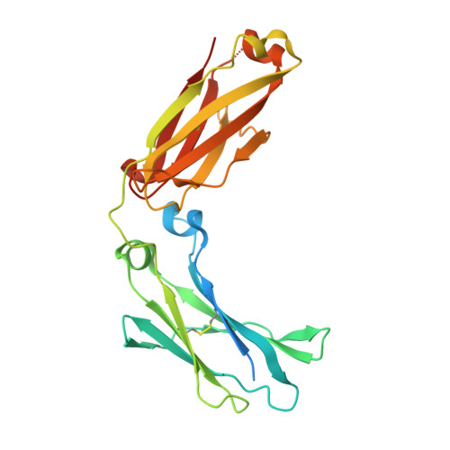
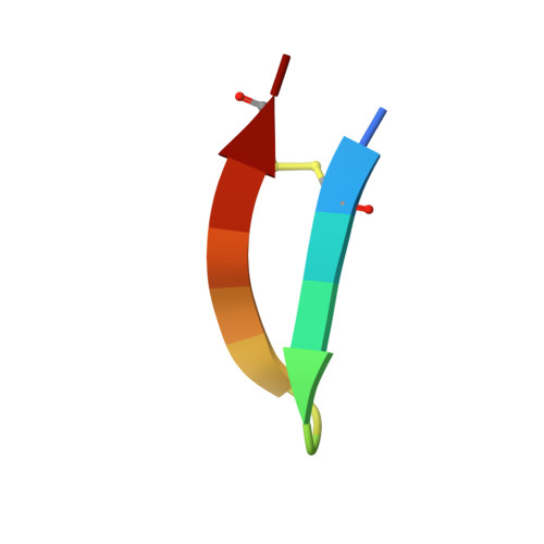
Computationally Designed Bispecific Antibodies using Negative State Repertoires.

Leaver-Fay, A.Froning, K.J.Atwell, S.Aldaz, H.Pustilnik, A.Lu, F.Huang, F.Yuan, R.Hassanali, S.Chamberlain, A.K.Fitchett, J.R.Demarest, S.J.Kuhlman, B.

(2016) Structure 24: 641-651

  • DOI: https://doi.org/10.1016/j.str.2016.02.013
  • Primary Citation Related Structures: 
    5DI8, 5DJ0, 5DJ2, 5DJ6, 5DJ8, 5DJA, 5DJC, 5DJD, 5DJX, 5DJY, 5DJZ, 5DK0, 5DK2, 5DVK, 5DVL, 5DVM, 5DVN, 5DVO

  • PubMed Abstract: 

    A challenge in the structure-based design of specificity is modeling the negative states, i.e., the complexes that you do not want to form. This is a difficult problem because mutations predicted to destabilize the negative state might be accommodated by small conformational rearrangements. To overcome this challenge, we employ an iterative strategy that cycles between sequence design and protein docking in order to build up an ensemble of alternative negative state conformations for use in specificity prediction. We have applied our technique to the design of heterodimeric CH3 interfaces in the Fc region of antibodies. Combining computationally and rationally designed mutations produced unique designs with heterodimer purities greater than 90%. Asymmetric Fc crystallization was able to resolve the interface mutations; the heterodimer structures confirmed that the interfaces formed as designed. With these CH3 mutations, and those made at the heavy-/light-chain interface, we demonstrate one-step synthesis of four fully IgG-bispecific antibodies.


  • Organizational Affiliation
    • Department of Biochemistry, University of North Carolina at Chapel Hill, 120 Mason Farm Road, Campus Box 7260, Chapel Hill, NC 27599, USA.

Macromolecule Content 

  • Total Structure Weight: 113.89 kDa 
  • Atom Count: 7,227 
  • Modeled Residue Count: 853 
  • Deposited Residue Count: 960 
  • Unique protein chains: 3

Macromolecules

Find similar proteins by:|  3D Structure
Entity ID: 1
MoleculeChains  Sequence LengthOrganismDetailsImage
Ig gamma-1 chain C region
A, D
227Homo sapiensMutation(s): 4 
Gene Names: IGHG1
UniProt & NIH Common Fund Data Resources
Find proteins for P01857 (Homo sapiens)
Explore P01857 
Go to UniProtKB:  P01857
PHAROS:  P01857
GTEx:  ENSG00000211896 
Entity Groups
Sequence Clusters30% Identity50% Identity70% Identity90% Identity95% Identity100% Identity
UniProt GroupP01857
Glycosylation
Glycosylation Sites: 1Go to GlyGen: P01857-2
Sequence Annotations
Expand
Reference Sequence
Find similar proteins by:|  3D Structure
Entity ID: 2
MoleculeChains  Sequence LengthOrganismDetailsImage
Ig gamma-1 chain C region
B, E
240Homo sapiensMutation(s): 7 
Gene Names: IGHG1
UniProt & NIH Common Fund Data Resources
Find proteins for P01857 (Homo sapiens)
Explore P01857 
Go to UniProtKB:  P01857
PHAROS:  P01857
GTEx:  ENSG00000211896 
Entity Groups
Sequence Clusters30% Identity50% Identity70% Identity90% Identity95% Identity100% Identity
UniProt GroupP01857
Glycosylation
Glycosylation Sites: 1Go to GlyGen: P01857-2
Sequence Annotations
Expand
Reference Sequence
Find similar proteins by:  Sequence   |   3D Structure  
Entity ID: 3
MoleculeChains  Sequence LengthOrganismDetailsImage
Fc-III peptide
C, F
13synthetic constructMutation(s): 0 

Oligosaccharides

Help  
Entity ID: 4
MoleculeChains Length2D Diagram GlycosylationD Interactions
beta-D-galactopyranose-(1-4)-2-acetamido-2-deoxy-beta-D-glucopyranose-(1-2)-alpha-D-mannopyranose-(1-6)-[alpha-D-mannopyranose-(1-3)]beta-D-mannopyranose-(1-4)-2-acetamido-2-deoxy-beta-D-glucopyranose-(1-4)-[alpha-L-fucopyranose-(1-6)]2-acetamido-2-deoxy-beta-D-glucopyranose
G, H, I, J
8N-Glycosylation
Glycosylation Resources
GlyTouCan: G61334IA
GlyCosmos: G61334IA
GlyGen: G61334IA

Small Molecules

Ligands 1 Unique
IDChains Name / Formula / InChI Key2D Diagram3D Interactions
IOD

Query on IOD


Download:Ideal Coordinates CCD File
K [auth D]IODIDE ION
I
XMBWDFGMSWQBCA-UHFFFAOYSA-M

Experimental Data & Validation

Experimental Data

  • Method: X-RAY DIFFRACTION
  • Resolution: 2.10 Å
  • R-Value Free:  0.285 (Depositor), 0.287 (DCC) 
  • R-Value Work:  0.229 (Depositor) 
  • R-Value Observed: 0.232 (Depositor) 
Space Group: P 1
Unit Cell:
Length ( Å )Angle ( ˚ )
a = 60.242α = 85.17
b = 66.858β = 80.71
c = 72.771γ = 89.69
Software Package:
Software NamePurpose
REFMACrefinement
SCALAdata scaling
PDB_EXTRACTdata extraction
iMOSFLMdata reduction
MOLREPphasing

Structure Validation

View Full Validation Report


Entry History 

Revision History  (Full details and data files)

  • Version 1.0: 2016-03-30
    Type: Initial release
  • Version 1.1: 2016-04-27
    Changes: Database references
  • Version 2.0: 2020-07-29
    Type: Remediation
    Reason: Carbohydrate remediation
    Changes: Advisory, Atomic model, Data collection, Database references, Derived calculations, Structure summary
  • Version 2.1: 2024-10-23
    Changes: Advisory, Data collection, Database references, Derived calculations, Structure summary