5DHD | pdb_00005dhd

Crystal structure of ChBD2 from Thermococcus kodakarensis KOD1


Experimental Data Snapshot

  • Method: X-RAY DIFFRACTION
  • Resolution: 1.27 Å
  • R-Value Free: 
    0.195 (Depositor), 0.214 (DCC) 
  • R-Value Work: 
    0.195 (Depositor), 0.214 (DCC) 

Starting Model: experimental
View more details

wwPDB Validation 3D Report Full Report

Validation slider image for 5DHD

This is version 1.2 of the entry. See complete history

Literature

Crystal structures of chitin binding domains of chitinase from Thermococcus kodakarensis KOD1

Hanazono, Y.Takeda, K.Niwa, S.Hibi, M.Takahashi, N.Kanai, T.Atomi, H.Miki, K.

(2016) FEBS Lett 590: 298-304

  • DOI: https://doi.org/10.1002/1873-3468.12055
  • Primary Citation Related Structures: 
    5DHD, 5DHE

  • PubMed Abstract: 

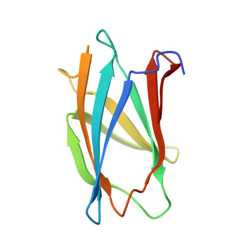
    Chitinase from T. kodakarensis (TkChiA) catalyzes the hydrolysis of chitin. The enzyme consists of two catalytic and three binding domains (ChBD1, ChBD2 and ChBD3). ChBD2 and ChBD3 can bind to not only chitin but also cellulose. In both domains, the intervals of the side chains of the three tryptophan residues, which are located on the molecular surface, correspond to twice the length of the lattice of the chitin. A binding model with crystalline chitin implies that the tryptophan residues and a glutamate residue interact with the hexose ring by CH-π interactions and the amide group by a hydrogen bond, respectively.


  • Organizational Affiliation
    • Department of Chemistry, Graduate School of Science, Kyoto University, Sakyo-ku, Japan.

Macromolecule Content 

  • Total Structure Weight: 14.93 kDa 
  • Atom Count: 959 
  • Modeled Residue Count: 98 
  • Deposited Residue Count: 100 
  • Unique protein chains: 1

Macromolecules

Find similar proteins by:|  3D Structure
Entity ID: 1
MoleculeChains  Sequence LengthOrganismDetailsImage
Chitinase100Thermococcus kodakarensis KOD1Mutation(s): 0 
Gene Names: Pk-chiATK1765
EC: 3.2.1.14
UniProt
Find proteins for Q9UWR7 (Thermococcus kodakarensis (strain ATCC BAA-918 / JCM 12380 / KOD1))
Explore Q9UWR7 
Go to UniProtKB:  Q9UWR7
Entity Groups
Sequence Clusters30% Identity50% Identity70% Identity90% Identity95% Identity100% Identity
UniProt GroupQ9UWR7
Sequence Annotations
Expand
Reference Sequence

Small Molecules

Ligands 2 Unique
IDChains Name / Formula / InChI Key2D Diagram3D Interactions
PE3

Query on PE3


Download:Ideal Coordinates CCD File
D [auth A]
E [auth A]
F [auth A]
G [auth A]
H [auth A]
D [auth A],
E [auth A],
F [auth A],
G [auth A],
H [auth A],
I [auth A]
3,6,9,12,15,18,21,24,27,30,33,36,39-TRIDECAOXAHENTETRACONTANE-1,41-DIOL
C28 H58 O15
ILLKMACMBHTSHP-UHFFFAOYSA-N
SO4

Query on SO4


Download:Ideal Coordinates CCD File
B [auth A],
C [auth A]
SULFATE ION
O4 S
QAOWNCQODCNURD-UHFFFAOYSA-L

Experimental Data & Validation

Experimental Data

  • Method: X-RAY DIFFRACTION
  • Resolution: 1.27 Å
  • R-Value Free:  0.195 (Depositor), 0.214 (DCC) 
  • R-Value Work:  0.195 (Depositor), 0.214 (DCC) 
Space Group: P 65
Unit Cell:
Length ( Å )Angle ( ˚ )
a = 60.245α = 90
b = 60.245β = 90
c = 68.845γ = 120
Software Package:
Software NamePurpose
CNSrefinement
DENZOdata collection
SCALEPACKdata scaling
MOLREPphasing
PDB_EXTRACTdata extraction

Structure Validation

View Full Validation Report


Entry History 

& Funding Information

Deposition Data


Funding OrganizationLocationGrant Number
CREST, JSTJapan--

Revision History  (Full details and data files)

  • Version 1.0: 2016-02-10
    Type: Initial release
  • Version 1.1: 2020-02-19
    Changes: Data collection, Database references, Derived calculations
  • Version 1.2: 2023-11-08
    Changes: Data collection, Database references, Refinement description