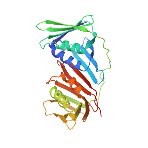
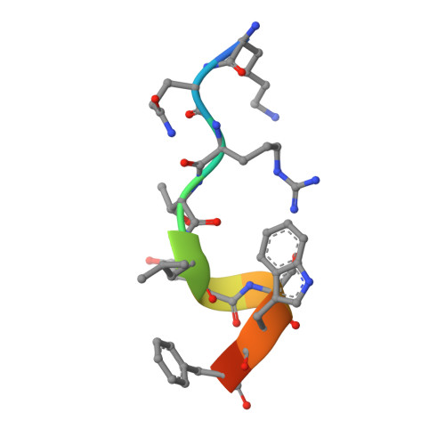
5DAI | pdb_00005dai

Proliferating cell nuclear antigen homolog 1 bound to FEN-1 peptide


Experimental Data Snapshot

  • Method: X-RAY DIFFRACTION
  • Resolution: 2.00 Å
  • R-Value Free: 
    0.224 (Depositor), 0.226 (DCC) 
  • R-Value Work: 
    0.181 (Depositor), 0.185 (DCC) 
  • R-Value Observed: 
    0.183 (Depositor) 

Starting Model: experimental
View more details

wwPDB Validation 3D Report Full Report

Validation slider image for 5DAI

This is version 1.3 of the entry. See complete history. 

Literature

A small protein inhibits proliferating cell nuclear antigen by breaking the DNA clamp.

Altieri, A.S., Ladner, J.E., Li, Z., Robinson, H., Sallman, Z.F., Marino, J.P., Kelman, Z.

(2016) Nucleic Acids Res 44: 6232-6241

  • DOI: https://doi.org/10.1093/nar/gkw351
  • Primary Citation Related Structures: 
    5DA7, 5DAI

  • PubMed Abstract: 

    Proliferating cell nuclear antigen (PCNA) forms a trimeric ring that encircles duplex DNA and acts as an anchor for a number of proteins involved in DNA metabolic processes. PCNA has two structurally similar domains (I and II) linked by a long loop (inter-domain connector loop, IDCL) on the outside of each monomer of the trimeric structure that makes up the DNA clamp. All proteins that bind to PCNA do so via a PCNA-interacting peptide (PIP) motif that binds near the IDCL. A small protein, called TIP, binds to PCNA and inhibits PCNA-dependent activities although it does not contain a canonical PIP motif. The X-ray crystal structure of TIP bound to PCNA reveals that TIP binds to the canonical PIP interaction site, but also extends beyond it through a helix that relocates the IDCL. TIP alters the relationship between domains I and II within the PCNA monomer such that the trimeric ring structure is broken, while the individual domains largely retain their native structure. Small angle X-ray scattering (SAXS) confirms the disruption of the PCNA trimer upon addition of the TIP protein in solution and together with the X-ray crystal data, provides a structural basis for the mechanism of PCNA inhibition by TIP.


  • Organizational Affiliation: 
    • Institute for Bioscience and Biotechnology Research, University of Maryland and the National Institute of Standards and Technology, 9600 Gudelsky Drive, Rockville, MD 20850, USA.

Macromolecule Content 

  • Total Structure Weight: 31.57 kDa 
  • Atom Count: 2,272 
  • Modeled Residue Count: 256 
  • Deposited Residue Count: 266 
  • Unique protein chains: 2

Macromolecules

Find similar proteins by:|  3D Structure
Entity ID: 1
MoleculeChains  Sequence LengthOrganismDetailsImage
DNA polymerase sliding clamp 1255Thermococcus kodakarensis KOD1Mutation(s): 0 
Gene Names: pcn1, TK0535
UniProt
Find proteins for Q5JF32 (Thermococcus kodakarensis (strain ATCC BAA-918 / JCM 12380 / KOD1))
Explore Q5JF32 
Go to UniProtKB:  Q5JF32
Entity Groups
Sequence Clusters30% Identity50% Identity70% Identity90% Identity95% Identity100% Identity
UniProt GroupQ5JF32
Sequence Annotations
Expand
Reference Sequence
Find similar proteins by:  Sequence   |   3D Structure  
Entity ID: 2
MoleculeChains  Sequence LengthOrganismDetailsImage
C-terminus of FEN-1 protein11Thermococcus kodakarensis KOD1Mutation(s): 0 
EC: 3.1
UniProt
Find proteins for Q5JGN0 (Thermococcus kodakarensis (strain ATCC BAA-918 / JCM 12380 / KOD1))
Explore Q5JGN0 
Go to UniProtKB:  Q5JGN0
Entity Groups
Sequence Clusters30% Identity50% Identity70% Identity90% Identity95% Identity100% Identity
UniProt GroupQ5JGN0
Sequence Annotations
Expand
Reference Sequence

Small Molecules

Ligands 1 Unique
IDChains Name / Formula / InChI Key2D Diagram3D Interactions
SO4

Query on SO4


Download:Ideal Coordinates CCD File
C [auth A]
D [auth A]
E [auth A]
F [auth A]
G [auth A]
C [auth A],
D [auth A],
E [auth A],
F [auth A],
G [auth A],
H [auth A],
I [auth A],
J [auth A],
K [auth A],
L [auth A],
M [auth B]
SULFATE ION
O4 S
QAOWNCQODCNURD-UHFFFAOYSA-L

Experimental Data & Validation

Experimental Data

  • Method: X-RAY DIFFRACTION
  • Resolution: 2.00 Å
  • R-Value Free:  0.224 (Depositor), 0.226 (DCC) 
  • R-Value Work:  0.181 (Depositor), 0.185 (DCC) 
  • R-Value Observed: 0.183 (Depositor) 
Space Group: H 3
Unit Cell:
Length ( Å )Angle ( ˚ )
a = 152.07α = 90
b = 152.07β = 90
c = 35.24γ = 120
Software Package:
Software NamePurpose
PHENIXrefinement
CrystalCleardata reduction
CrystalCleardata scaling
PHASERphasing

Structure Validation

View Full Validation Report


Entry History 

Deposition Data

Revision History  (Full details and data files)

  • Version 1.0: 2016-05-11
    Type: Initial release
  • Version 1.1: 2016-05-18
    Changes: Database references
  • Version 1.2: 2016-08-10
    Changes: Database references
  • Version 1.3: 2023-09-27
    Changes: Data collection, Database references, Derived calculations, Refinement description