5DA5 | pdb_00005da5

Crystal structure of Rhodospirillum rubrum Rru_A0973


Experimental Data Snapshot

  • Method: X-RAY DIFFRACTION
  • Resolution: 2.06 Å
  • R-Value Free: 
    0.206 (Depositor), 0.203 (DCC) 
  • R-Value Work: 
    0.171 (Depositor), 0.167 (DCC) 
  • R-Value Observed: 
    0.173 (Depositor) 

Starting Model: experimental
View more details

wwPDB Validation 3D Report Full Report

Validation slider image for 5DA5

This is version 2.1 of the entry. See complete history

Literature

Structural characterization of encapsulated ferritin provides insight into iron storage in bacterial nanocompartments.

He, D.Hughes, S.Vanden-Hehir, S.Georgiev, A.Altenbach, K.Tarrant, E.J.Mackay, C.L.Waldron, K.J.Clarke, D.J.Marles-Wright, J.

(2016) Elife 5

  • DOI: https://doi.org/10.7554/eLife.18972
  • Primary Citation Related Structures: 
    5DA5, 5L89, 5L8B, 5L8G

  • PubMed Abstract: 

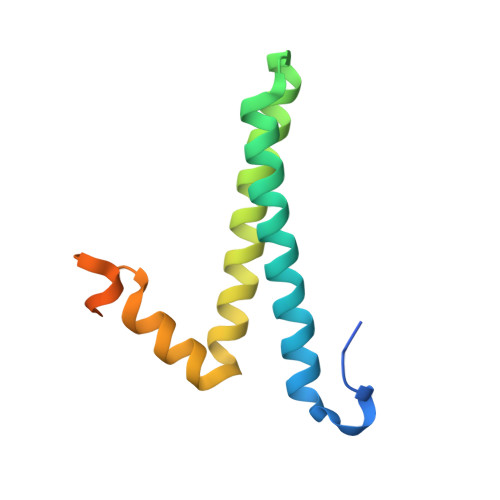
    Ferritins are ubiquitous proteins that oxidise and store iron within a protein shell to protect cells from oxidative damage. We have characterized the structure and function of a new member of the ferritin superfamily that is sequestered within an encapsulin capsid. We show that this encapsulated ferritin (EncFtn) has two main alpha helices, which assemble in a metal dependent manner to form a ferroxidase center at a dimer interface. EncFtn adopts an open decameric structure that is topologically distinct from other ferritins. While EncFtn acts as a ferroxidase, it cannot mineralize iron. Conversely, the encapsulin shell associates with iron, but is not enzymatically active, and we demonstrate that EncFtn must be housed within the encapsulin for iron storage. This encapsulin nanocompartment is widely distributed in bacteria and archaea and represents a distinct class of iron storage system, where the oxidation and mineralization of iron are distributed between two proteins.


  • Organizational Affiliation
    • Institute of Quantitative Biology, Biochemistry and Biotechnology, School of Biological Sciences, The University of Edinburgh, Edinburgh, United Kingdom.

Macromolecule Content 

  • Total Structure Weight: 404.44 kDa 
  • Atom Count: 23,222 
  • Modeled Residue Count: 2,703 
  • Deposited Residue Count: 3,480 
  • Unique protein chains: 1

Macromolecules

Find similar proteins by:|  3D Structure
Entity ID: 1
MoleculeChains  Sequence LengthOrganismDetailsImage
Rru_A0973116Rhodospirillum rubrumMutation(s): 0 
Gene Names: Rru_A0973
EC: 1.16.3.1
UniProt
Find proteins for Q2RVS1 (Rhodospirillum rubrum (strain ATCC 11170 / ATH 1.1.1 / DSM 467 / LMG 4362 / NCIMB 8255 / S1))
Explore Q2RVS1 
Go to UniProtKB:  Q2RVS1
Entity Groups
Sequence Clusters30% Identity50% Identity70% Identity90% Identity95% Identity100% Identity
UniProt GroupQ2RVS1
Sequence Annotations
Expand
Reference Sequence

Small Molecules

Ligands 3 Unique
IDChains Name / Formula / InChI Key2D Diagram3D Interactions
GOA

Query on GOA


Download:Ideal Coordinates CCD File
AB [auth G]
AC [auth Q]
FA [auth A]
FC [auth U]
HC [auth V]
AB [auth G],
AC [auth Q],
FA [auth A],
FC [auth U],
HC [auth V],
IB [auth K],
KB [auth L],
LA [auth B],
MA [auth C],
MC [auth X],
PA [auth D],
QB [auth N],
QC [auth Z],
VA [auth F],
VB [auth P],
XC [auth a]
GLYCOLIC ACID
C2 H4 O3
AEMRFAOFKBGASW-UHFFFAOYSA-N
FE

Query on FE


Download:Ideal Coordinates CCD File
CB [auth G]
CC [auth U]
EA [auth A]
EC [auth U]
FB [auth K]
CB [auth G],
CC [auth U],
EA [auth A],
EC [auth U],
FB [auth K],
GC [auth V],
HA [auth A],
HB [auth K],
IA [auth B],
JB [auth L],
KA [auth B],
KC [auth V],
LC [auth X],
OA [auth D],
OB [auth L],
OC [auth X],
PB [auth N],
PC [auth Z],
SA [auth D],
SB [auth N],
SC [auth Z],
TC [auth a],
UA [auth F],
UB [auth P],
WC [auth a],
XA [auth F],
XB [auth P],
YB [auth Q],
ZA [auth G],
ZB [auth Q]
FE (III) ION
Fe
VTLYFUHAOXGGBS-UHFFFAOYSA-N
CA

Query on CA


Download:Ideal Coordinates CCD File
BB [auth G]
BC [auth Q]
DB [auth I]
DC [auth U]
EB [auth J]
BB [auth G],
BC [auth Q],
DB [auth I],
DC [auth U],
EB [auth J],
GA [auth A],
GB [auth K],
IC [auth V],
JA [auth B],
JC [auth V],
LB [auth L],
MB [auth L],
NA [auth C],
NB [auth L],
NC [auth X],
QA [auth D],
RA [auth D],
RB [auth N],
RC [auth Z],
TA [auth D],
TB [auth N],
UC [auth a],
VC [auth a],
WA [auth F],
WB [auth P],
YA [auth F],
YC [auth a],
ZC [auth c]
CALCIUM ION
Ca
BHPQYMZQTOCNFJ-UHFFFAOYSA-N

Experimental Data & Validation

Experimental Data

  • Method: X-RAY DIFFRACTION
  • Resolution: 2.06 Å
  • R-Value Free:  0.206 (Depositor), 0.203 (DCC) 
  • R-Value Work:  0.171 (Depositor), 0.167 (DCC) 
  • R-Value Observed: 0.173 (Depositor) 
Space Group: P 1 21 1
Unit Cell:
Length ( Å )Angle ( ˚ )
a = 98.184α = 90
b = 120.533β = 95.36
c = 140.25γ = 90
Software Package:
Software NamePurpose
PHENIXrefinement
Aimlessdata scaling
XDSdata reduction
PHASERphasing

Structure Validation

View Full Validation Report


Entry History 

& Funding Information

Deposition Data


Funding OrganizationLocationGrant Number
China Scholarships CouncilChina--
Royal SocietyUnited KingdomRG130585

Revision History  (Full details and data files)

  • Version 1.0: 2016-08-10
    Type: Initial release
  • Version 1.1: 2016-08-31
    Changes: Database references
  • Version 1.2: 2018-01-31
    Changes: Author supporting evidence
  • Version 2.0: 2023-11-15
    Changes: Advisory, Atomic model, Data collection, Database references, Derived calculations
  • Version 2.1: 2024-01-10
    Changes: Refinement description