5D7V | pdb_00005d7v

Crystal structure of PTK6 kinase domain


Experimental Data Snapshot

  • Method: X-RAY DIFFRACTION
  • Resolution: 2.33 Å
  • R-Value Free: 
    0.282 (Depositor), 0.279 (DCC) 
  • R-Value Work: 
    0.219 (Depositor), 0.218 (DCC) 
  • R-Value Observed: 
    0.222 (Depositor) 

Starting Model: experimental
View more details

wwPDB Validation 3D Report Full Report

Validation slider image for 5D7V

This is version 1.3 of the entry. See complete history

Literature

Crystal structure of the kinase domain of human protein tyrosine kinase 6 (PTK6) at 2.33 angstrom resolution

Thakur, M.K.Kumar, A.Birudukota, S.Swaminathan, S.Tyagi, R.Gosu, R.

(2016) Biochem Biophys Res Commun 478: 637-642

  • DOI: https://doi.org/10.1016/j.bbrc.2016.07.121
  • Primary Citation Related Structures: 
    5D7V

  • PubMed Abstract: 

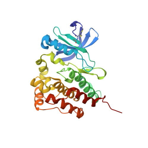
    Human Protein tyrosine kinase 6 (PTK6) (EC:2.7.10.2), also known as the breast tumor kinase (BRK), is an intracellular non-receptor Src-related tyrosine kinase expressed in a majority of human breast tumors and breast cancer cell lines, but its expression is low or completely absent in normal mammary glands. In the recent past, several studies have suggested that PTK6 is a potential therapeutic target in cancer. To understand its structural and functional properties, the PTK6 kinase domain (PTK6-KD) gene was cloned, overexpressed in a baculo-insect cell system, purified and crystallized at room temperature. X-ray diffraction data to 2.33 Å resolution was collected on a single PTK6-KD crystal, which belonged to the triclinic space group P1. The Matthews coefficient calculation suggested the presence of four protein molecules per asymmetric unit, with a solvent content of ∼50%.The structure has been solved by molecular replacement and crystal structure data submitted to the protein data bank under the accession number 5D7V. This is the first report of apo PTK6-KD structure crystallized in DFG-in and αC-helix-out conformation.


  • Organizational Affiliation
    • Department of Biochemistry, University of Mysore, Mysore, 570005, India; Department of Structural Biology, Jubilant Biosys Ltd, Bangalore, 560022, India.

Macromolecule Content 

  • Total Structure Weight: 126.05 kDa 
  • Atom Count: 9,141 
  • Modeled Residue Count: 1,064 
  • Deposited Residue Count: 1,076 
  • Unique protein chains: 1

Macromolecules

Find similar proteins by:|  3D Structure
Entity ID: 1
MoleculeChains  Sequence LengthOrganismDetailsImage
Protein-tyrosine kinase 6
A, B, C, D
269Homo sapiensMutation(s): 1 
Gene Names: PTK6
EC: 2.7.10.2
UniProt & NIH Common Fund Data Resources
Find proteins for Q13882 (Homo sapiens)
Explore Q13882 
Go to UniProtKB:  Q13882
PHAROS:  Q13882
GTEx:  ENSG00000101213 
Entity Groups
Sequence Clusters30% Identity50% Identity70% Identity90% Identity95% Identity100% Identity
UniProt GroupQ13882
Sequence Annotations
Expand
Reference Sequence

Small Molecules

Ligands 2 Unique
IDChains Name / Formula / InChI Key2D Diagram3D Interactions
PO4

Query on PO4


Download:Ideal Coordinates CCD File
E [auth A],
I [auth B],
M [auth C],
Q [auth D]
PHOSPHATE ION
O4 P
NBIIXXVUZAFLBC-UHFFFAOYSA-K
GOL

Query on GOL


Download:Ideal Coordinates CCD File
F [auth A]
G [auth A]
H [auth A]
J [auth B]
K [auth B]
F [auth A],
G [auth A],
H [auth A],
J [auth B],
K [auth B],
L [auth B],
N [auth C],
O [auth C],
P [auth C],
R [auth D],
S [auth D],
T [auth D]
GLYCEROL
C3 H8 O3
PEDCQBHIVMGVHV-UHFFFAOYSA-N
Modified Residues  1 Unique
IDChains TypeFormula2D DiagramParent
CXM
Query on CXM
A, B, C, D
L-PEPTIDE LINKINGC6 H11 N O4 SMET

Experimental Data & Validation

Experimental Data

  • Method: X-RAY DIFFRACTION
  • Resolution: 2.33 Å
  • R-Value Free:  0.282 (Depositor), 0.279 (DCC) 
  • R-Value Work:  0.219 (Depositor), 0.218 (DCC) 
  • R-Value Observed: 0.222 (Depositor) 
Space Group: P 1
Unit Cell:
Length ( Å )Angle ( ˚ )
a = 49.718α = 81.46
b = 76.308β = 75.92
c = 87.271γ = 74.93
Software Package:
Software NamePurpose
REFMACrefinement
PDB_EXTRACTdata extraction
HKL-2000data reduction
HKL-2000data scaling
MOLREPphasing

Structure Validation

View Full Validation Report


Entry History 

Deposition Data

Revision History  (Full details and data files)

  • Version 1.0: 2016-08-17
    Type: Initial release
  • Version 1.1: 2016-09-07
    Changes: Database references
  • Version 1.2: 2023-11-08
    Changes: Data collection, Database references, Derived calculations, Refinement description
  • Version 1.3: 2024-11-13
    Changes: Structure summary