5D45

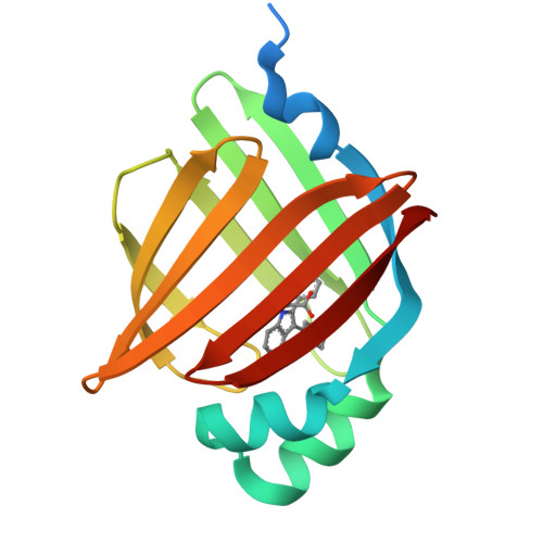
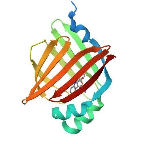
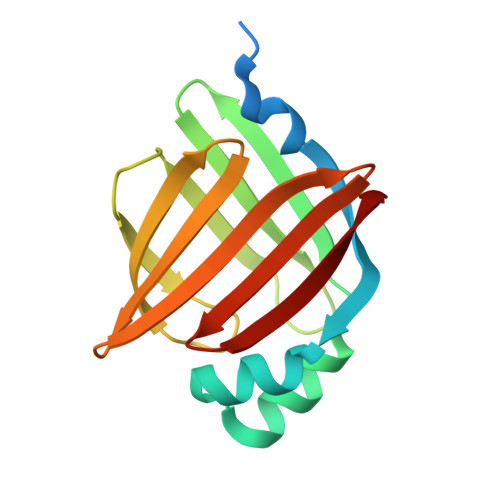
Crystal Structure of FABP4 in complex with 3-(5-cyclopropyl-2,3-diphenyl-1H-indol-1-yl)propanoic acid


Experimental Data Snapshot

  • Method: X-RAY DIFFRACTION
  • Resolution: 1.65 Å
  • R-Value Free: 0.217 
  • R-Value Work: 0.188 
  • R-Value Observed: 0.189 

Starting Model: experimental
View more details

wwPDB Validation   3D Report Full Report


Ligand Structure Quality Assessment 


This is version 1.2 of the entry. See complete history


Literature

Interaction Analysis of FABP4 Inhibitors by X-ray Crystallography and Fragment Molecular Orbital Analysis

Tagami, U.Takahashi, K.Igarashi, S.Ejima, C.Yoshida, T.Takeshita, S.Miyanaga, W.Sugiki, M.Tokumasu, M.Hatanaka, T.Kashiwagi, T.Ishikawa, K.Miyano, H.Mizukoshi, T.

(2016) ACS Med Chem Lett 7: 435-439

  • DOI: https://doi.org/10.1021/acsmedchemlett.6b00040
  • Primary Citation of Related Structures:  
    5D45, 5D47, 5D48, 5D4A

  • PubMed Abstract: 

    X-ray crystal structural determination of FABP4 in complex with four inhibitors revealed the complex binding modes, and the resulting observations led to improvement of the inhibitory potency of FABP4 inhibitors. However, the detailed structure-activity relationship (SAR) could not be explained from these structural observations. For a more detailed understanding of the interactions between FABP4 and inhibitors, fragment molecular orbital analyses were performed. These analyses revealed that the total interfragment interaction energies of FABP4 and each inhibitor correlated with the ranking of the K i value for the four inhibitors. Furthermore, interactions between each inhibitor and amino acid residues in FABP4 were identified. The oxygen atom of Lys58 in FABP4 was found to be very important for strong interactions with FABP4. These results might provide useful information for the development of novel potent FABP4 inhibitors.


  • Organizational Affiliation

    Institute for Innovation, Ajinomoto Co., Inc. , 1-1 Suzuki-cho, Kawasaki 210-8681, Japan.


Macromolecules
Find similar proteins by:  (by identity cutoff)  |  3D Structure
Entity ID: 1
MoleculeChains Sequence LengthOrganismDetailsImage
Fatty acid-binding protein, adipocyte152Homo sapiensMutation(s): 0 
Gene Names: FABP4
UniProt & NIH Common Fund Data Resources
Find proteins for P15090 (Homo sapiens)
Explore P15090 
Go to UniProtKB:  P15090
PHAROS:  P15090
GTEx:  ENSG00000170323 
Entity Groups  
Sequence Clusters30% Identity50% Identity70% Identity90% Identity95% Identity100% Identity
UniProt GroupP15090
Sequence Annotations
Expand
  • Reference Sequence
Small Molecules
Ligands 1 Unique
IDChains Name / Formula / InChI Key2D Diagram3D Interactions
57P
Query on 57P

Download Ideal Coordinates CCD File 
B [auth A]3-(5-cyclopropyl-2,3-diphenyl-1H-indol-1-yl)propanoic acid
C26 H23 N O2
VTCPSZSCIMRYND-UHFFFAOYSA-N
Binding Affinity Annotations 
IDSourceBinding Affinity
57P BindingDB:  5D45 Ki: 120 (nM) from 1 assay(s)
Experimental Data & Validation

Experimental Data

  • Method: X-RAY DIFFRACTION
  • Resolution: 1.65 Å
  • R-Value Free: 0.217 
  • R-Value Work: 0.188 
  • R-Value Observed: 0.189 
  • Space Group: P 21 21 21
Unit Cell:
Length ( Å )Angle ( ˚ )
a = 32.39α = 90
b = 54.16β = 90
c = 74.6γ = 90
Software Package:
Software NamePurpose
XSCALEdata scaling
MOLREPphasing
REFMACrefinement
PDB_EXTRACTdata extraction

Structure Validation

View Full Validation Report


Ligand Structure Quality Assessment 


Entry History 

Revision History  (Full details and data files)

  • Version 1.0: 2016-06-22
    Type: Initial release
  • Version 1.1: 2020-02-19
    Changes: Data collection, Derived calculations
  • Version 1.2: 2023-11-08
    Changes: Data collection, Database references, Refinement description