


Structure of CYP107L2 from Streptomyces avermitilis with lauric acid
Pham, T.-V., Han, S.-H., Kim, J.-H., Kim, D.-H., Kang, L.-W.To be published.
Experimental Data Snapshot
Starting Model: experimental
View more details
Entity ID: 1 | |||||
|---|---|---|---|---|---|
| Molecule | Chains | Sequence Length | Organism | Details | Image |
| Cytochrome P450 hydroxylase | 393 | Streptomyces avermitilis MA-4680 = NBRC 14893 | Mutation(s): 0 Gene Names: cyp8, SAV_1987 | 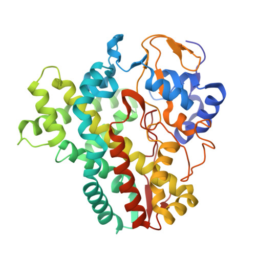 | |
UniProt | |||||
Entity Groups | |||||
| Sequence Clusters | 30% Identity50% Identity70% Identity90% Identity95% Identity100% Identity | ||||
| UniProt Group | Q82LM3 | ||||
Sequence AnnotationsExpand | |||||
Reference Sequence | |||||
| Ligands 4 Unique | |||||
|---|---|---|---|---|---|
| ID | Chains | Name / Formula / InChI Key | 2D Diagram | 3D Interactions | |
| HEM Download:Ideal Coordinates CCD File | E [auth A], H [auth B] | PROTOPORPHYRIN IX CONTAINING FE C34 H32 Fe N4 O4 KABFMIBPWCXCRK-RGGAHWMASA-L |  | ||
| DAO Download:Ideal Coordinates CCD File | D [auth A], G [auth B] | LAURIC ACID C12 H24 O2 POULHZVOKOAJMA-UHFFFAOYSA-N |  | ||
| SO4 Download:Ideal Coordinates CCD File | C [auth A], F [auth B] | SULFATE ION O4 S QAOWNCQODCNURD-UHFFFAOYSA-L |  | ||
| GOL Download:Ideal Coordinates CCD File | I [auth B] | GLYCEROL C3 H8 O3 PEDCQBHIVMGVHV-UHFFFAOYSA-N |  | ||
| Length ( Å ) | Angle ( ˚ ) |
|---|---|
| a = 64.08 | α = 90 |
| b = 118.371 | β = 105.51 |
| c = 66.039 | γ = 90 |
| Software Name | Purpose |
|---|---|
| REFMAC | refinement |
| DENZO | data processing |
| MOLREP | model building |
| PHASER | phasing |
| HKL-2000 | data processing |
| Coot | model building |


