5CVR | pdb_00005cvr

Crystal structure of FNR of A. fischeri in a partially degraded form


Experimental Data Snapshot

  • Method: X-RAY DIFFRACTION
  • Resolution: 2.60 Å
  • R-Value Free: 
    0.237 (Depositor), 0.259 (DCC) 
  • R-Value Work: 
    0.191 (Depositor), 0.209 (DCC) 
  • R-Value Observed: 
    0.193 (Depositor) 

Starting Model: other
View more details

wwPDB Validation 3D Report Full Report

Validation slider image for 5CVR

This is version 1.2 of the entry. See complete history

Literature

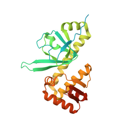
The crystal structure of the global anaerobic transcriptional regulator FNR explains its extremely fine-tuned monomer-dimer equilibrium.

Volbeda, A.Darnault, C.Renoux, O.Nicolet, Y.Fontecilla-Camps, J.C.

(2015) Sci Adv 1: e1501086-e1501086

  • DOI: https://doi.org/10.1126/sciadv.1501086
  • Primary Citation Related Structures: 
    5CVR, 5E44

  • PubMed Abstract: 

    The structure of the dimeric holo-fumarate and nitrate reduction regulator (FNR) from Aliivibrio fischeri has been solved at 2.65 Å resolution. FNR globally controls the transition between anaerobic and aerobic respiration in facultative anaerobes through the assembly/degradation of its oxygen-sensitive [4Fe-4S] cluster. In the absence of O2, FNR forms a dimer and specifically binds to DNA, whereas in its presence, the cluster is degraded causing FNR monomerization and DNA dissociation. We have used our crystal structure and the information previously gathered from numerous FNR variants to propose that this process is governed by extremely fine-tuned interactions, mediated by two salt bridges near the amino-terminal cluster-binding domain and an "imperfect" coiled-coil dimer interface. [4Fe-4S] to [2Fe-2S] cluster degradation propagates a conformational signal that indirectly causes monomerization by disrupting the first of these interactions and unleashing the "unzipping" of the FNR dimer in the direction of the carboxyl-terminal DNA binding domain.


  • Organizational Affiliation
    • Metalloproteins Unit, Institut de Biologie Structurale, CEA, CNRS, Université Grenoble Alpes, 71, Avenue des Martyrs CS10090, 38044 Grenoble, France.

Macromolecule Content 

  • Total Structure Weight: 29.75 kDa 
  • Atom Count: 1,626 
  • Modeled Residue Count: 206 
  • Deposited Residue Count: 259 
  • Unique protein chains: 1

Macromolecules

Find similar proteins by:|  3D Structure
Entity ID: 1
MoleculeChains  Sequence LengthOrganismDetailsImage
FNR type regulator259Aliivibrio fischeriMutation(s): 0 
Gene Names: fnr
UniProt
Find proteins for Q70ET4 (Aliivibrio fischeri)
Explore Q70ET4 
Go to UniProtKB:  Q70ET4
Entity Groups
Sequence Clusters30% Identity50% Identity70% Identity90% Identity95% Identity100% Identity
UniProt GroupQ70ET4
Sequence Annotations
Expand
Reference Sequence

Experimental Data & Validation

Experimental Data

  • Method: X-RAY DIFFRACTION
  • Resolution: 2.60 Å
  • R-Value Free:  0.237 (Depositor), 0.259 (DCC) 
  • R-Value Work:  0.191 (Depositor), 0.209 (DCC) 
  • R-Value Observed: 0.193 (Depositor) 
Space Group: I 4 2 2
Unit Cell:
Length ( Å )Angle ( ˚ )
a = 75.27α = 90
b = 75.27β = 90
c = 212.43γ = 90
Software Package:
Software NamePurpose
Aimlessdata scaling
PHENIXrefinement
PDB_EXTRACTdata extraction
PHENIXphasing
XDSdata reduction

Structure Validation

View Full Validation Report


Entry History 

Deposition Data

Revision History  (Full details and data files)

  • Version 1.0: 2015-12-16
    Type: Initial release
  • Version 1.1: 2015-12-23
    Changes: Database references
  • Version 1.2: 2024-05-01
    Changes: Data collection, Database references, Refinement description