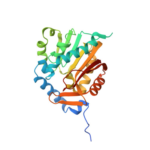
5CVE

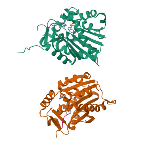
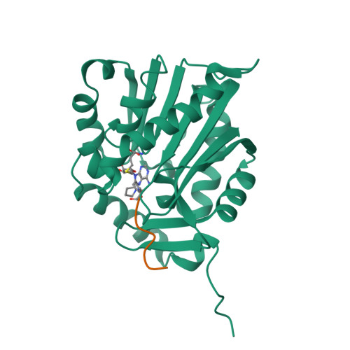
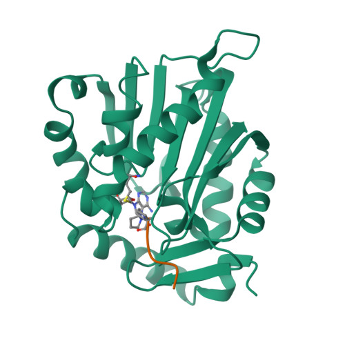
Crystal Structure of human NRMT1 in complex with dimethylated fly H2B peptide and SAH


Experimental Data Snapshot

  • Method: X-RAY DIFFRACTION
  • Resolution: 1.50 Å
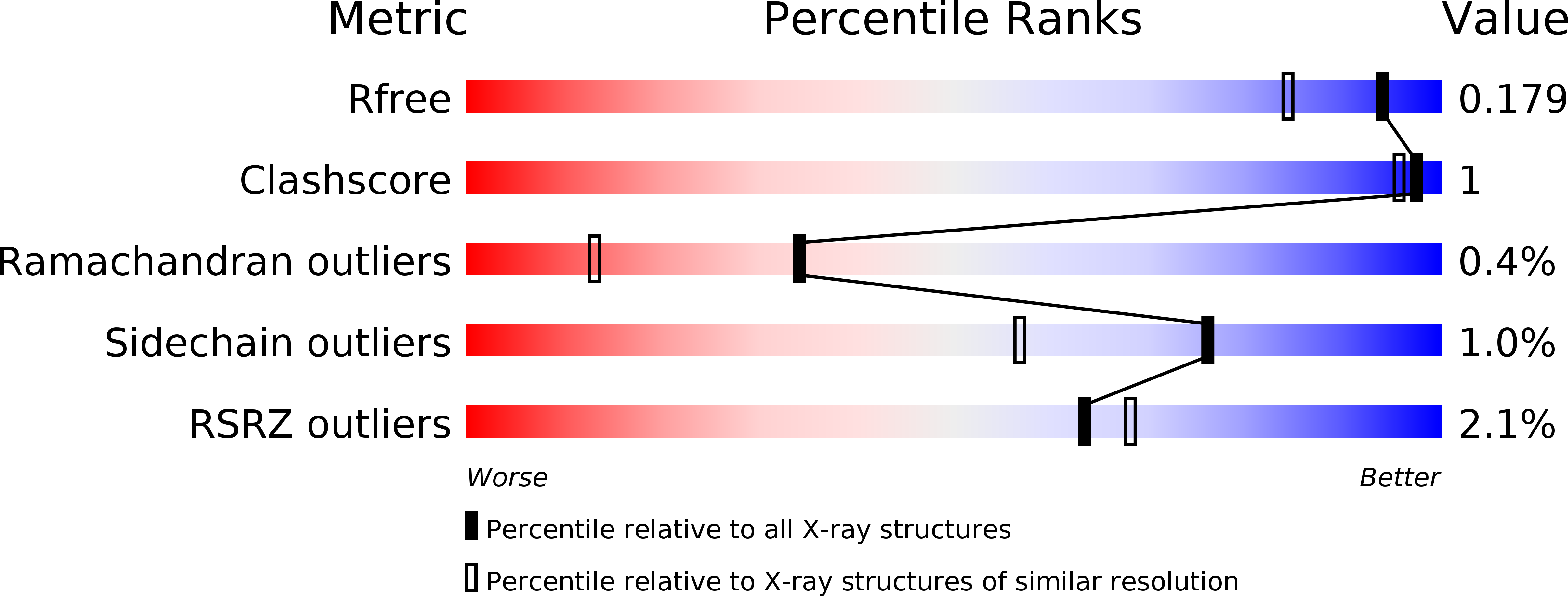
  • R-Value Free: 0.179 
  • R-Value Work: 0.153 
  • R-Value Observed: 0.153 

wwPDB Validation   3D Report Full Report


Ligand Structure Quality Assessment 


This is version 1.2 of the entry. See complete history


Literature

Molecular basis for histone N-terminal methylation by NRMT1

Wu, R.Yue, Y.Zheng, X.Li, H.

(2015) Genes Dev 29: 2337-2342

  • DOI: https://doi.org/10.1101/gad.270926.115
  • Primary Citation of Related Structures:  
    5CVD, 5CVE

  • PubMed Abstract: 

    NRMT1 is an N-terminal methyltransferase that methylates histone CENP-A as well as nonhistone substrates. Here, we report the crystal structure of human NRMT1 bound to CENP-A peptide at 1.3 Å. NRMT1 adopts a core methyltransferase fold that resembles DOT1L and PRMT but not SET domain family histone methyltransferases. Key substrate recognition and catalytic residues were identified by mutagenesis studies. Histone peptide profiling revealed that human NRMT1 is highly selective to human CENP-A and fruit fly H2B, which share a common "Xaa-Pro-Lys/Arg" motif. These results, along with a 1.5 Å costructure of human NRMT1 bound to the fruit fly H2B peptide, underscore the importance of the NRMT1 recognition motif.


  • Organizational Affiliation

    MOE Key Laboratory of Protein Sciences, Center for Structural Biology, Department of Basic Medical Sciences, School of Medicine, Tsinghua University, Beijing 100084, China;


Macromolecules
Find similar proteins by:  (by identity cutoff)  |  3D Structure
Entity ID: 1
MoleculeChains Sequence LengthOrganismDetailsImage
N-terminal Xaa-Pro-Lys N-methyltransferase 1
A, B
243Homo sapiensMutation(s): 0 
Gene Names: NTMT1NRMT1
EC: 2.1.1.244
UniProt & NIH Common Fund Data Resources
Find proteins for Q9BV86 (Homo sapiens)
Explore Q9BV86 
Go to UniProtKB:  Q9BV86
PHAROS:  Q9BV86
GTEx:  ENSG00000148335 
Entity Groups  
Sequence Clusters30% Identity50% Identity70% Identity90% Identity95% Identity100% Identity
UniProt GroupQ9BV86
Sequence Annotations
Expand
  • Reference Sequence

Find similar proteins by:  Sequence   |   3D Structure  

Entity ID: 2
MoleculeChains Sequence LengthOrganismDetailsImage
N-terminal peptide from Histone H2BC [auth D],
D [auth E]
9Drosophila melanogasterMutation(s): 0 
UniProt
Find proteins for P02283 (Drosophila melanogaster)
Explore P02283 
Go to UniProtKB:  P02283
Entity Groups  
UniProt GroupP02283
Sequence Annotations
Expand
  • Reference Sequence
Small Molecules
Ligands 1 Unique
IDChains Name / Formula / InChI Key2D Diagram3D Interactions
SAH
Query on SAH

Download Ideal Coordinates CCD File 
E [auth A],
F [auth B]
S-ADENOSYL-L-HOMOCYSTEINE
C14 H20 N6 O5 S
ZJUKTBDSGOFHSH-WFMPWKQPSA-N
Experimental Data & Validation

Experimental Data

  • Method: X-RAY DIFFRACTION
  • Resolution: 1.50 Å
  • R-Value Free: 0.179 
  • R-Value Work: 0.153 
  • R-Value Observed: 0.153 
  • Space Group: C 1 2 1
Unit Cell:
Length ( Å )Angle ( ˚ )
a = 114.836α = 90
b = 66.302β = 106.44
c = 68.73γ = 90
Software Package:
Software NamePurpose
HKL-2000data collection
HKL-2000data scaling
PHENIXrefinement
PDB_EXTRACTdata extraction
SCALAdata reduction

Structure Validation

View Full Validation Report


Ligand Structure Quality Assessment 


Entry History 

Deposition Data

  • Released Date: 2015-11-25 
  • Deposition Author(s): Wu, R., Li, H.

Revision History  (Full details and data files)

  • Version 1.0: 2015-11-25
    Type: Initial release
  • Version 1.1: 2015-12-02
    Changes: Database references
  • Version 1.2: 2017-09-27
    Changes: Data collection, Database references, Derived calculations