


Crystal structure of Trypanosoma cruzi protein in complex with ligand
Yang, Y.Y., Ko, T.P., Zheng, Y.Y., Liu, W.D., Chen, C.C., Guo, R.T.(2016) ACS Chem Biol
Experimental Data Snapshot
wwPDB Validation 3D Report Full Report

(2016) ACS Chem Biol
Entity ID: 1 | |||||
|---|---|---|---|---|---|
| Molecule | Chains | Sequence Length | Organism | Details | Image |
| Acidocalcisomal pyrophosphatase | 414 | Trypanosoma cruzi | Mutation(s): 0 EC: 3.6.1.1 | 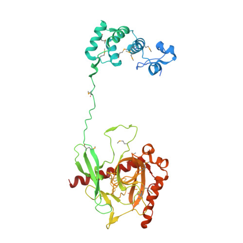 | |
UniProt | |||||
Entity Groups | |||||
| Sequence Clusters | 30% Identity50% Identity70% Identity90% Identity95% Identity100% Identity | ||||
| UniProt Group | Q4JH30 | ||||
Sequence AnnotationsExpand | |||||
Reference Sequence | |||||
| Ligands 2 Unique | |||||
|---|---|---|---|---|---|
| ID | Chains | Name / Formula / InChI Key | 2D Diagram | 3D Interactions | |
| MLT Download:Ideal Coordinates CCD File | C [auth A] | D-MALATE C4 H6 O5 BJEPYKJPYRNKOW-UWTATZPHSA-N |  | ||
| MG Download:Ideal Coordinates CCD File | D [auth A], E [auth B] | MAGNESIUM ION Mg JLVVSXFLKOJNIY-UHFFFAOYSA-N |  | ||
| Modified Residues 1 Unique | |||||
|---|---|---|---|---|---|
| ID | Chains | Type | Formula | 2D Diagram | Parent |
| MSE Query on MSE | A, B | L-PEPTIDE LINKING | C5 H11 N O2 Se |  | MET |
| Length ( Å ) | Angle ( ˚ ) |
|---|---|
| a = 100.518 | α = 90 |
| b = 103.017 | β = 90 |
| c = 157.398 | γ = 90 |
| Software Name | Purpose |
|---|---|
| HKL-2000 | data scaling |
| CNS | refinement |
| PDB_EXTRACT | data extraction |
| HKL-2000 | data reduction |
| CNS | phasing |


